Let’s start with the most common misunderstanding: the place that hurts is usually not the place that’s broken.
Almost everyone with a trigger finger points to their middle knuckle and says, “It gets stuck here.” But the real problem is at the base of the palm, that ring where the finger meets the palm. This is why the doctor presses your palm, not the spot you’re pointing at.
This is written for those whose fingers get stuck in the morning, who feel pain when gripping things, or who have already been told they need an injection.
What exactly is this?
For your fingers to bend, they rely on a few tendons that run all the way from the forearm to the fingertips. The tendons don’t flop around because there are a series of ring-like “pulleys” along the way holding them tight against the bones, much like the line guides on a fishing rod.
The pulley closest to the palm has the highest chance of causing trouble.
When the tendon swells up, or the pulley gets thicker, their sizes no longer match. When you bend your finger, the flexor muscles are strong enough to force the swollen part through. But when you try to straighten it, the extensor muscles are too weak to pull it back.
And so the finger gets stuck in a bent position.
That “snap” sound is the swollen part finally squeezing through the pulley.
By the way, the formal name for this condition is “stenosing tenosynovitis,” but it’s a bit of a misnomer—under a microscope, the changes are mainly in the pulley itself and its surrounding tissue, not inflammation of the synovium. This detail will matter later when we get to “why injections stop working if you wait too long.”
The four grades that decide what you should do
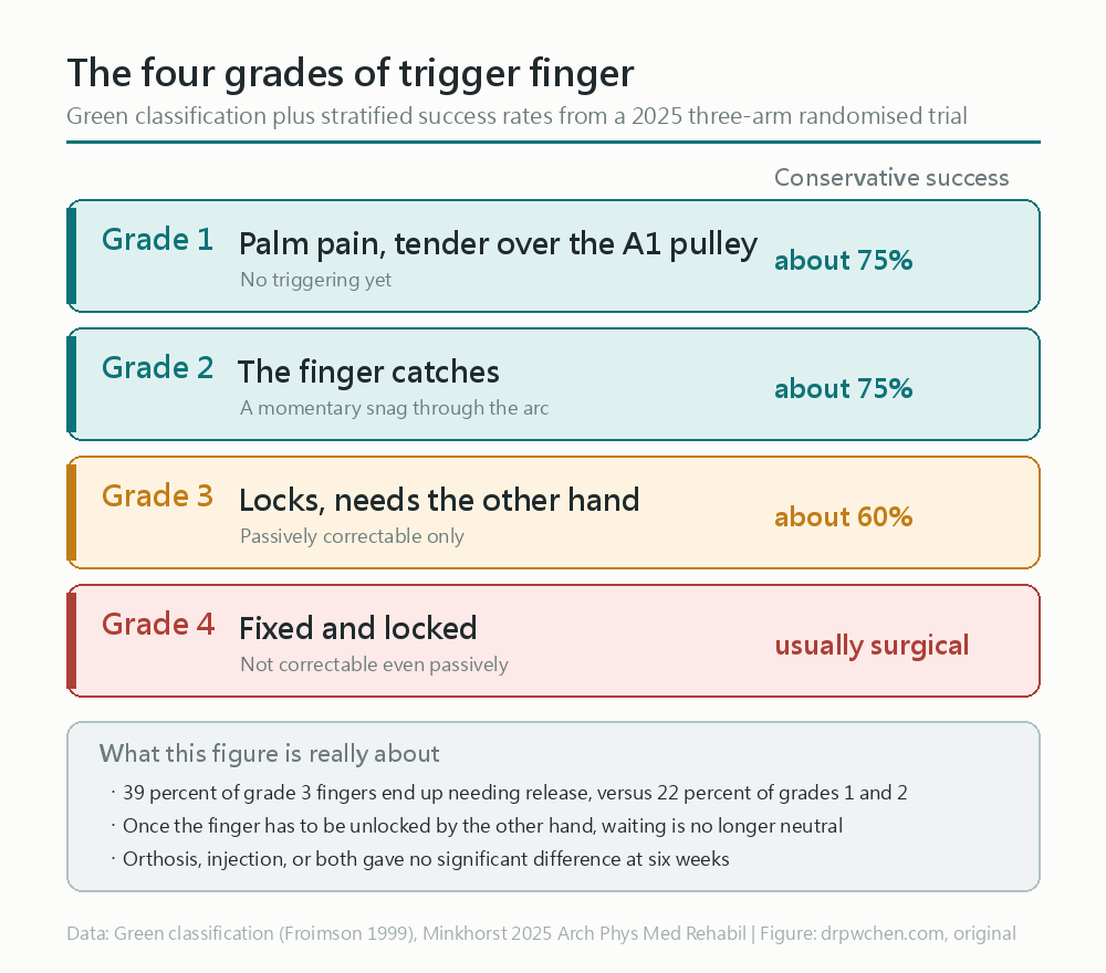
The most important takeaway from this chart is this: by the time you have to pry it open with your other hand, the situation is already quite different.
A 2025 randomized controlled trial tracked 122 fingers, and the results were: for people in grades one and two, conservative treatment (splinting, injections, or both) works about three-quarters of the time. But by grade three, the success rate drops to 60%, and the rate of ultimately needing surgery jumps from 22% to 39%.
So if you are already using your other hand to pry your finger open, you’re not in the “let’s wait and see” stage anymore.
This is a problem that snowballs on its own
Here’s something worth knowing: trigger finger gets worse on its own, not because you aren’t being careful enough.
Every time it gets stuck, the tendon gets rubbed. Every time it gets rubbed, it swells a little more. When it swells more, it gets stuck more easily. This cycle keeps spinning on its own.
So the strategy of “just putting up with it to see if it heals on its own” doesn’t really work for trigger finger. It might get better on its own, but it usually takes several months.
Who is more likely to get it?
- Diabetes — this is the most important one. About 2% of the general population gets trigger finger, but the rate is much higher for people with diabetes (different studies put it anywhere from 7% to 20%).
- Women, significantly more so than men.
- Peaking between ages 40 and 60.
- Rheumatoid arthritis, gout, hypothyroidism.
- Long-term repetitive gripping with the hand.
But let’s be clear: for most people, there is no obvious cause. It’s not necessarily because they “overused their hands.” Very few actually have a history of trauma.
There’s another one that’s easily overlooked: people who have had carpal tunnel surgery have a higher chance of developing a trigger finger afterward. This is a confirmed link, not a coincidence.
As for which finger gets it most often, many people think it’s the middle or ring finger. In reality, the thumb is number one (about one-third), followed by the ring finger. Kids are completely different—90% of pediatric trigger fingers are in the thumb, and a quarter of them have it in both hands.
Splints, injections, or surgery: how do you choose?
Splints
The logic is simple: immobilize the joint where the finger meets the palm, so the tendon stops sliding back and forth through that pulley, giving the inflamed area a chance to rest.
The joints lower down the finger need to stay mobile so it doesn’t mess up your daily life.
How well it works is highly dependent on “how long you’ve put it off”: if symptoms have been around for less than six months, the success rate is about 77%; within four months, it’s about 73%. If you wait too long, or if multiple fingers are involved, it doesn’t work as well.
Splints are less effective for trigger thumbs. Every report agrees on this.
Injections
Injecting steroids into that spot on the palm is the most common approach.
I need to clear up a common misconception: this injection doesn’t go into the joint. It goes into or around the tendon sheath, outside the tendon. When the doctor gives the shot, they’ll ask you to wiggle your finger—that’s just to confirm the needle hasn’t pierced the tendon itself.
How well does it work?
- After a single injection, about 54% to 73% of people are symptom-free a year later.
- Repeated injections are safe and have a compounding effect, but the longer you wait, the more shots you’ll need.
- Afterward, you’re usually told to wear a splint or protect it for a week.
- It might hurt quite a bit for a few days after the shot. This is expected; they didn’t botch it.
One thing worth noting: even though ultrasound guidance is more precise, current studies don’t show better clinical outcomes with it. So if your doctor doesn’t use ultrasound, it doesn’t mean they’re being careless.
When should you consider surgery?
- Two injections didn’t fix it.
- You’re already at grade four (completely locked).
- People with poorly controlled diabetes might want to discuss this sooner.
That third point is worth expanding on. A study looked at 297 patients and found that for those with an HbA1c over 6.5%, the injection failure rate was 71.9%, compared to 38.1% for those with good control.
That’s nearly double the difference.
The same study found two other predictors of failure: having another hand condition on the same hand (like De Quervain’s or carpal tunnel), and having had symptoms for more than two and a half months.
Surgery itself has a very high success rate, between 90% and 100%. But it’s not zero-risk. Large-scale statistics show minor complications around 10% and major complications around 2.4%, with persistent stiffness, swelling, and pain being the most common. So it’s “a good option after conservative treatment fails,” not “cut early to save trouble.”
Why injections stop working if you wait too long
This is the part I think is most worth talking about.
I mentioned earlier that the microscopic changes aren’t just inflammation. Instead, the tissue has already started altering into a “cartilage-like” state.
Steroids can suppress inflammation, but they can’t undo tissue that has already changed character.
So “why injections stop working the more you get them” isn’t because your body is resistant to steroids, nor is it a problem with the doctor’s technique. It’s because the disease process has already reached a stage where medication can’t handle it. This is why dealing with it early versus waiting until later yields such vastly different results.
Do painkillers help?
Oral or topical NSAIDs can be used for initial symptom control.
But if we’re talking about “getting an NSAID injection instead of a steroid injection,” a 2021 Cochrane review compared them: there was no significant difference in symptom resolution rates, but a higher proportion of those who got NSAID injections still had moderate-to-severe symptoms later on (28% versus 14%).
The conclusion is that there is no reason to replace steroids with NSAID injections.
When should you see a doctor?
- Your finger requires prying open with your other hand to straighten it.
- You can’t straighten it at all, and it won’t pry open either.
- You’ve had two injections but keep having flare-ups.
- You have diabetes, and your blood sugar control is sub-optimal.
- Your finger is red, swollen, hot, and very painful to the touch (this requires ruling out an infection, it’s not just a simple trigger finger).
That last one is the most important. Trigger finger itself doesn’t make your finger red and hot. If it is, you’ve got something else going on, and you need to get it checked out ASAP.
If the thumb side of your wrist is also hurting, that might be a different condition. You can check out De Quervain’s tenosynovitis.
Clinical Pearls
- Trigger finger is a size mismatch between the flexor tendon and the A1 pulley (at the MCP joint level), causing triggering or locking in flexion
- The lesion is at the A1 pulley, but patients mostly point to the PIP joint for pain; look for tenderness and nodules near the distal palmar crease
- Resisted movements do not provoke pain (to distinguish from flexor tendinitis)
- First-line treatment is corticosteroid injection into/around the A1 pulley sheath, not intra-articular; the overall success rate for conservative management is about 70%
- Failure rate for injections is 71.9% in those with HbA1c > 6.5% (vs 38.1%); early surgical consultation is advised
- Histologically, it’s fibrocartilaginous metaplasia of the retinacular sheath, which explains why steroids lose efficacy later in the disease course
Abbreviations
| Abbreviation | Full Name |
|---|---|
| A1 pulley | First annular pulley |
| FDS | Flexor digitorum superficialis |
| FDP | Flexor digitorum profundus |
| MCP | Metacarpophalangeal joint |
| PIP | Proximal interphalangeal joint |
| DIP | Distal interphalangeal joint |
| CTS | Carpal tunnel syndrome |
| DM | Diabetes mellitus |
| RA | Rheumatoid arthritis |
| ROM | Range of motion |
| PRWHE | Patient-Reported Wrist and Hand Evaluation |
Disease Background
Definition
- When the flexor tendon catches, snaps, or locks as it glides through the A1 pulley, that’s a trigger finger
Cyriax Ch.73,Frontera 4e Ch.37 - Full name: stenosing flexor tenosynovitis
- “Tenosynovitis” is actually a misnomer: the histopathological changes are located in the retinacular sheath and peritendinous tissue, not the tenosynovium.
Tenovaginitisis the more accurate termFrontera 4e Ch.37
- “Tenosynovitis” is actually a misnomer: the histopathological changes are located in the retinacular sheath and peritendinous tissue, not the tenosynovium.
- Core pathology: thickening of the retinacular sheath + disproportionate narrowing of the caliber relative to the tendon, stemming from hypertrophy and fibrocartilaginous metaplasia at the tendon–pulley interface
- Why it catches at the A1 pulley: this is where the tendon enters the pulley system at the steepest angle, making pressure highest at the proximal edge
- Mechanical explanation
Cyriax Ch.73- Flexors are strong enough to pull the nodule past the resistance (flexion can still be completed)
- Extensors are weaker and cannot pull the nodule back proximal to the pulley → finger locked in flexion
Two Presentations
| Type | Lesion Distribution | Palpation |
|---|---|---|
| Nodular stenosing tenosynovitis | Thickening localized to a short segment distal to the A1 pulley | Distinct nodule is palpable |
| Diffuse stenosing tenosynovitis | Inflammation extends along the tendon, beyond the A1 pulley | No discrete nodule is palpable |
(Magee Pathology 2e Ch.10)
Epidemiology
- Prevalence in the general population is about 2%
Frontera 4e Ch.37,JAMA 2022 - The proportion in the diabetic population is much higher than in the general population, though the figures from two sources vary widely and should be presented together:
- The older series cited by Frontera is 7%
Frontera 4e Ch.37 - The JAMA 2022 review notes that up to 20% of adults with diabetes are affected
JAMA 2022
- The older series cited by Frontera is 7%
- Peak age is 40–60 years
Kahn Ch.31; Cyriax’s series notes 50–60 yearsCyriax Ch.73 - More common in women than men; the Cyriax series states it can be up to 6 times more common
Cyriax Ch.73 - The distribution of affected fingers is completely different between adults and children
Frontera 4e Ch.37- Adults: Thumb 33%, ring finger 27% are the top two (Magee’s order: thumb > ring > middle > little > index)
Magee Pathology 2e Ch.10 - Children: 90% are in the thumb, of which 25% are bilateral
- Adults: Thumb 33%, ring finger 27% are the top two (Magee’s order: thumb > ring > middle > little > index)
Risk Factors and Comorbidities
- Systemic
Kahn Ch.31,Frontera 4e Ch.37- Diabetes mellitus (most important, affects treatment response)
- Rheumatoid arthritis (can present as a secondary trigger finger)
- Gout, hypothyroidism, amyloidosis, histiocytosis, other connective tissue diseases
- Local
- Repetitive forceful gripping; however, most cases have no clear trigger and are idiopathic
Magee Pathology 2e Ch.10 - A new-onset trigger digit after carpal tunnel release is a recognized association
Magee Pathology 2e Ch.10 - Those with concurrent lesions in the ipsilateral hand have a higher injection failure rate
Hollins 2022
- Repetitive forceful gripping; however, most cases have no clear trigger and are idiopathic
- A clear history of trauma is actually rare; causation by acute trauma or space-occupying lesions is uncommon
Kahn Ch.31,Frontera 4e Ch.37
Natural History
- It is a self-reinforcing vicious cycle: repetitive friction from triggering continues to exacerbate tendon swelling → triggering worsens
Kahn Ch.31 - Typical diurnal variation: stiffness and locking are most pronounced in the morning or after prolonged inactivity, gradually loosening up with movement
- Spontaneous resolution can occur, but takes months
Cyriax Ch.24
Prognosis
- Overall success rate of conservative treatment (orthosis/injection/both) is 68.9%
Minkhorst 2025- Stratified by Green’s classification: Grade 1–2 is about 75%, Grade 3 drops to 60%
- The proportion of Grade 3 ultimately undergoing surgical release is 39.1% vs 22.4% for lower grades (P = .05)
- After a single A1 pulley injection, the 1-year symptom resolution rate is 54–73%
Frontera 4e Ch.37 - Surgical (open A1 pulley release) success rate is 90–100%
Elahi 2023 - Three predictors of injection failure
Hollins 2022:- HbA1c > 6.5%: failure rate 71.9% vs 38.1% (P < 0.001)
- Concurrent hand pathology in the same hand: 43.4% vs 30.8% (P = 0.032)
- Symptoms lasting over 2.5 months: 40.4% vs 29.5% (P = 0.048)
- Injection efficacy is poorer for a disease course longer than 4 months or when multiple fingers are involved simultaneously
Frontera 4e Ch.37- Mechanistic explanation: steroids cannot reverse established fibrocartilaginous metaplasia
Complications
- Disease itself
Frontera 4e Ch.37- PIP joint flexion contracture (most common permanent sequela)
- Rare: intractable pain persisting after treatment
- Injection-related
- Skin depigmentation, dermatitis, subcutaneous fat atrophy, finger necrosis, tendon rupture, injury to sensory branches of digital nerves, infection
- Post-injection after-pain can be quite significant
Cyriax Ch.24
- Surgery-related
Elahi 2023- Large series (543 patients/795 digits): minor complications 9.6%, major 2.4%
- Small single-surgeon series (43 patients/78 digits): minor up to 28%, major 3% (synovial fistula, PIP arthrofibrosis)
- Most common are persistent stiffness, swelling, and pain; increased risk with sedation, general anesthesia, and male sex
- Specific risks: bowstringing (excessive release), unresolved triggering (incomplete release), radial digital nerve injury (especially watch out in the thumb, as this nerve crosses directly over the A1 pulley)
Clinical Assessment
Diagnostic Criteria
- Purely a clinical diagnosis: history + physical examination is sufficient
Kahn Ch.31,Frontera 4e Ch.37 - Routine blood work is not needed; order relevant tests only if undiagnosed DM/RA/gout/amyloidosis is suspected
- Routine X-rays are not needed (if no history of trauma or inflammatory arthritis); order only when suspecting sesamoid abnormalities, MCP loose bodies, metacarpal head osteophytes, or collateral ligament avulsion
Patient History
- Early stage: intermittent pain in the palm/finger + swelling + triggering
- Progression: digit locks in flexion, requiring passive extension with the other hand; snap + pain at the moment of extension
- The chief complaint of pain can be deceiving: as narrowing worsens, patients often complain of PIP joint pain, while the true lesion is at the MCP joint level
Frontera 4e Ch.37 - Trigger thumb and trigger finger present differently: the thumb primarily presents with pain during motion, whereas other fingers primarily present with triggering and loss of ROM
Frontera 4e Ch.37 - Three things to ask: when does triggering occur (is it during gripping?), how long have symptoms lasted, and are they progressing?
Kahn Ch.31 - Resisted movements do not provoke pain (important for distinguishing from flexor tendinitis)
Cyriax Ch.24
Physical Examination
- Palpable tender nodule: located around the distal palmar crease, i.e., at the A1 pulley level; for trigger thumb, it’s proximal to the 1st metacarpal head
- Have the patient flex and extend their finger while palpating the nodule; you can feel it move under your finger, and a click may be heard/felt
- Make sure to check the passive ROM of all finger joints (MCP/PIP/DIP): true triggering will also catch during passive movement
Kahn Ch.31 - Retest active flexion and extension to see if it can clear the catching point
- Chronic cases may present with an IP joint flexion contracture
- Neurological exam should be normal (unless complicated by CTS/diabetic neuropathy, or severe disuse weakness)
Imaging
- Imaging is not used for diagnosis, but can serve as an adjunct
Frontera 4e Ch.37- MRI can confirm flexor sheath tenosynovitis, but offers virtually no additional value over clinical diagnosis
- Ultrasound can reveal tendon nodules and sheath effusions, and allows for dynamic observation of the actual catching at the A1 pulley level
- Ultrasound findings
Malanga Ch.40- A1 pulley = a thin hypoechoic line volar to the flexor tendon, located at the MCP joint and slightly proximal to it
- Tenosynovitis = a hypoechoic layer around the tendon + enlargement of the tendon and sheath distal to the A1 pulley
- Use Doppler to locate the digital arteries and nerves on both sides first before planning the needle trajectory
- Anatomic localization (surface landmarks)
Malanga Ch.40- The A1 pulley originates from the MCP joint volar plate, is located about 5 mm proximal to the joint, and is about 1 cm long
- Using the proximal finger crease as a surface landmark: the proximal edge of the pulley is about 2 cm away, and the distal edge is about 1 cm away
Severity Grading
Two grading systems exist simultaneously; clinical notes must specify which one is being used.
Green classification (established by Froimson in 1999, most commonly used clinically and in recent RCTs) Kahn Ch.31, Minkhorst 2025
| Grade | Presentation |
|---|---|
| I | Pain in the palm, tenderness at the A1 pulley (no triggering yet) |
| II | Finger catching |
| III | Finger locking, passively correctable |
| IV | Fixed and locked, uncorrectable |
Quinnell classification (1980, adopted by Frontera, includes an additional type 0) Frontera 4e Ch.37
| Type | Presentation |
|---|---|
| 0 | Crepitus only, no triggering |
| I | Uneven movement but no triggering |
| II | Triggering is actively correctable |
| III | Triggering is only passively correctable |
| IV | Fixed deformity |
Differential Diagnosis
| Category | DDx | Key Distinguishing Features |
|---|---|---|
| Tendon | Flexor tendinitis | Pain on resisted flexion; no triggering nodule |
| Tendon | Flexor / extensor tendon rupture | Loss of active movement rather than catching |
| Tendon | Extensor digitorum communis subluxation | Snapping is on the dorsal side, not at the volar A1 pulley |
| Fascia | Dupuytren’s contracture | Contracted cords in the palmar aponeurosis; no snapping; flexion contracture of the ring/little finger |
| Joint | Finger joint arthritis | Capsular pattern of restriction; no triggering |
| Joint | Chronic MCP dislocation / loose body / abnormal sesamoid | Visible on imaging; catching point during passive movement doesn’t match the A1 pulley |
| Ligament | Sagittal band rupture | Extensor tendon subluxation during finger extension |
| Tumor | Giant cell tumor of tendon sheath, ganglion, amyloid deposits | Mass does not move with flexion and extension; identifiable on imaging |
| Systemic | Rheumatoid arthritis | RA can cause secondary trigger finger; requires overall systemic assessment |
| Systemic | Acromegaly | Acromegalic features; multiple digits simultaneously involved |
| Infection | Tendon sheath infection | Erythema, swelling, heat, Kanavel’s four cardinal signs |
(Cyriax Ch.73, Kahn Ch.31, Frontera 4e Ch.37)
Clinical Management
Treatment Algorithm
Management algorithm
Consensus framework (European HANDGUIDE Delphi, 35 hand surgeons / hand therapists / physiatrists, achieved over 4 rounds)
HANDGUIDE 2014
- Four endorsed treatment options: orthosis, corticosteroid injection, injection + orthosis combined, surgery
- Choice is based on three factors: severity, disease duration, prior treatments received
Practical pathway
- Grade I–II or short disease duration → orthosis + activity modification + (if necessary) NSAIDs
- Persistent symptoms or functional limitation → corticosteroid injection into/around the A1 pulley sheath
- Unresolved after two injections → refer to hand surgery
- Diabetes (especially insulin-dependent, HbA1c > 6.5%), multiple digits involved, recurrences → consider early surgery
Cost-effectiveness perspective: Non-invasive treatment → max two injections → surgery, is the recommended ladder
Frontera 4e Ch.37
Orthoses
- Goal: Limit tendon excursion through the A1 pulley, reduce friction, and let the inflamed tissue rest
Webster Atlas 5e Ch.16 - Common prescription: MCP immobilized in 0°–15° of flexion, with PIP and DIP left free, worn continuously for up to 6 weeks
Frontera 4e Ch.37,Kahn Ch.31- Differences in schools of thought: Evans advocates for MCP in full extension; Patel/Eaton advocate for 10–15° of flexion for better patient tolerance
Webster Atlas 5e Ch.16 - Leaving the IP joints free allows for a hook fist, maintaining function while limiting full tendon glide
- Differences in schools of thought: Evans advocates for MCP in full extension; Patel/Eaton advocate for 10–15° of flexion for better patient tolerance
- Success rate (observational data, not RCTs)
Webster Atlas 5e Ch.16- Symptoms under 6 months: 77%; symptoms under 4 months: 73%
- Efficacy is poorer for thumbs and when multiple digits are involved
- An alternative approach immobilizing the DIP joint reports long-term success rates up to 87%, though similarly poorer in the thumb
Frontera 4e Ch.37
- Orthoses can reduce days lost from work
Frontera 4e Ch.37 - Adding an orthosis to an injection offers no additional benefit: a 3-arm RCT (orthosis vs injection vs combined, 122 digits) showed no significant differences between the three groups at 6 weeks in the number of triggerings, symptom relief, or PRWHE scores
Minkhorst 2025
Pharmacotherapy
- Oral/topical NSAIDs: can be used for initial symptom control
Frontera 4e Ch.37 - NSAID injections cannot replace corticosteroid injections: Two Cochrane RCTs (231 participants) compared a single NSAID injection vs corticosteroid injection
Cochrane 2021- No difference in symptom resolution rates (NSAID 34% vs steroid 41%, RR 0.83, 95% CI 0.62–1.11, low certainty)
- However, more patients in the NSAID group had persistent moderate-to-severe symptoms (28% vs 14%, RR 2.03, 95% CI 1.19–3.46)
- Conclusion: NSAID injections offer little to no benefit relative to steroids
Injection Therapy
- Location is intrasheath or perisheath, not intra-articular
- Must never be injected into the tendon substance: if the needle moves when the patient is asked to flex their finger after insertion, it means you’re in the tendon, and you must withdraw slightly
Kahn Ch.31
- Must never be injected into the tendon substance: if the needle moves when the patient is asked to flex their finger after insertion, it means you’re in the tendon, and you must withdraw slightly
- Medications and equipment
- Cyriax technique: triamcinolone acetonide 10 mg / 1 mL, 1 mL tuberculin syringe, finest 2 cm needle
Cyriax Ch.24 - Frontera technique: 27G 5/8 inch needle, 1 mL 1% lidocaine + 1 mL (40 mg) methylprednisolone, inserted at the distal palmar crease
Frontera 4e Ch.37 - Common under ultrasound guidance: 25G 1 inch needle, 10 mg Depo-Medrone + 0.5 mL 1% lidocaine, small hockey stick probe
Resteghini Ch.7.4 - It is currently unclear which steroid is superior (betamethasone is water-soluble, has a lower risk of tenosynovitis and fat necrosis, and is often recommended, but lacks evidence of clinical superiority)
Frontera 4e Ch.37
- Cyriax technique: triamcinolone acetonide 10 mg / 1 mL, 1 mL tuberculin syringe, finest 2 cm needle
- Technical pearls
- Trigger thumb: supine, palm up; needle inserted 1 cm distal to the nodule, directed proximally; half injected around the nodule, half injected inside it; review at 2 weeks, very rarely needs a second one
Cyriax Ch.24 - Other fingers: A1 pulley (metacarpal head) level, needle at 30° to the skin
Cyriax Ch.73 - Ultrasound short-axis out-of-plane approach, must absolutely avoid the digital arteries on both sides
Narouze Ch.21
- Trigger thumb: supine, palm up; needle inserted 1 cm distal to the nodule, directed proximally; half injected around the nodule, half injected inside it; review at 2 weeks, very rarely needs a second one
- Efficacy
- Cochrane: steroid + lidocaine is superior to lidocaine alone, treatment success at 4 weeks RR 3.15 (95% CI 1.34–7.40), NNT of 3
Cochrane 2009- Low-quality evidence: only 2 small RCTs (63 participants total) with poor methodological quality, rated silver level by Cochrane; in one trial, efficacy was maintained up to 4 months
- 1-year symptom resolution after a single injection is 54–73%; repeated injections are safe and have an additive effect, but the longer the disease course, the more injections needed
Frontera 4e Ch.37 - Ultrasound guidance has not been shown to be clinically superior to landmark-guided injection (though helpful for localization and anatomic variations)
Frontera 4e Ch.37 - Routine post-injection practice: orthosis and relative protection for 1 week
- Cochrane: steroid + lidocaine is superior to lidocaine alone, treatment success at 4 weeks RR 3.15 (95% CI 1.34–7.40), NNT of 3
- Other injectates: intrasheath hyaluronic acid and diclofenac both have reports of clinical benefit, but evidence volume is low
Frontera 4e Ch.37
Deep Transverse Friction
- Indication: flexor pollicis longus tenosynovitis at the carpal level (when crepitus is present)
Cyriax Ch.24 - DTF is ineffective for trigger thumb at the MCP level; it requires an injection
Rehabilitation
- Goal: Maintain ROM, prevent tendon adhesions and contractures
Frontera 4e Ch.37,Kahn Ch.31 - Scenarios appropriate for occupational/physical therapy referral:
- Loss of strength/ROM/function due to disuse or prolonged immobilization
- Need for a custom orthosis (better fit and occupational functionality than off-the-shelf)
- Need for modalities to reduce inflammation (ice massage, contrast baths, paraffin wax, ultrasound, iontophoresis)
- Both pre- and post-operative supervised therapy can improve ROM, strength, and function
- Padded gloves can prevent direct pressure on the palm
Surgical Intervention
- Indications
Kahn Ch.31,Frontera 4e Ch.37- Failure of conservative treatment, unresolved after two injections
- Suspicion of other pathology such as RA causing the triggering
- Patient desires rapid and definitive relief
- Populations more likely to need surgery: diabetes, RA, multiple joint involvement, younger age of onset
- Two surgical techniques
- Open A1 pulley release (gold standard): Under local anesthesia, make a 1–1.5 cm incision on the volar aspect at the MCP joint level (following skin creases to minimize scarring), longitudinally incise the A1 pulley at least to the A2 pulley level; ask the patient to flex and extend on the spot to confirm complete release
Elahi 2023 - Percutaneous release: Percutaneous incision using a needle; a meta-analysis reports success rates up to 94%, with better results under ultrasound guidance
Frontera 4e Ch.37 - An RCT (Gilberts et al.) showed no difference in recurrence rates between open and percutaneous release; however, open offers better visualization and may be safer for avoiding iatrogenic neurovascular injury
Elahi 2023
- Open A1 pulley release (gold standard): Under local anesthesia, make a 1–1.5 cm incision on the volar aspect at the MCP joint level (following skin creases to minimize scarring), longitudinally incise the A1 pulley at least to the A2 pulley level; ask the patient to flex and extend on the spot to confirm complete release
- The catching site is the A1 pulley in almost all cases, but a minority are at A2, A3, or the palmar aponeurosis
Elahi 2023 - Post-operative
- Most patients can resume activity immediately post-op; common complaint is soreness in the palm
- Early active and passive ROM, 4 times daily
- Recovery is mostly within weeks, but swelling and stiffness may take 6 months to fully resolve
- Still lacking full ROM at the first post-op visit → refer to formal physical therapy
Return to Work / Sports
- May resume daily activities 3–7 days post-injection (no formal guidelines; peak analgesic effect may take days to weeks, but this is a comfort issue and does not restrict return to play)
Kahn Ch.31 - Return to activity is mostly immediate post-surgery
Indications for Referral
- Unresolved after two injections, or Grade III–IV
- Poorly controlled diabetes, multiple digit involvement, or recurrence → early referral
- Post-op emergence of: infection, radial digital nerve injury, incomplete extension (distal sheath still tight), unresolved triggering (incomplete release), bowstringing (over-release)
How These Notes Were Generated
These notes were originally my go-to reference for hand problems in the clinic. I’ve completely overhauled them this time to post on the site.
Materials used
- PM&R and orthopedic textbooks: Frontera Essentials of PM&R 4e, Ombregt (Cyriax) 3e, Kahn Musculoskeletal Sports and Spine Disorders, Magee Pathology and Intervention 2e, Webster Atlas of Orthoses and Assistive Devices 5e
- Ultrasound-guided injection textbooks: Malanga, Resteghini, Narouze
- Society consensus: European HANDGUIDE multidisciplinary Delphi consensus guideline (Phys Ther 2014)
- Primary literature: Two Cochrane systematic reviews (corticosteroid injections 2009, NSAID injections 2021), JAMA 2022 review of common hand conditions, a 2025 3-arm RCT (orthosis vs injection vs combined), a retrospective study on HbA1c and injection failure, and a case series on open surgery complications
Tools used
audit_note.py— A note format auditing script I wrote to check citation placement, images, and heading structure- PubMed MCP — To find society consensus and systematic reviews, and to fetch missing volume/issue/page numbers, DOIs, and PMIDs for the bibliography
- textbook_search — For semantic search across the local markdown indices of my textbooks
- OpenEvidence — For an independent cross-search to verify existing conclusions
- My own Python/PIL drawing script — For the grading diagram and cover image (
scripts/figures/) - Hugo’s bilingual workflow
i18n_sync.py— Machine translating the English version first, then manually proofreading the medical terms
This whole workflow is written up in textbook-to-note. If you have a Claude or Codex subscription, you can farm out all this mechanical labor of verification and compilation to it, leaving the judgment calls to yourself. You can start with Getting started with AI: installation and first steps and How to talk to AI agents.
About this version
The professional section is pulled directly from my own clinical notes. It hasn’t been rewritten for the blog, and the citation format follows my note-taking convention (Book Title + Chapter, or Author + Year, with the full bibliography below). Copyrighted images from textbooks and journals are not included here; all diagrams were redrawn by myself.
If there are any errors in my understanding, corrections are more than welcome.
Reference
Guidelines / Consensus
- HANDGUIDE 2014 — Huisstede BMA, Hoogvliet P, Coert JH, Fridén J. Multidisciplinary consensus guideline for managing trigger finger: results from the European HANDGUIDE Study. Phys Ther. 2014;94(10):1421-1433. doi:10.2522/ptj.20130135. PMID 24810861
Journals
- Peters-Veluthamaningal C, van der Windt DAWM, Winters JC, Meyboom-de Jong B. Corticosteroid injection for trigger finger in adults. Cochrane Database Syst Rev. 2009;(1):CD005617. doi:10.1002/14651858.CD005617.pub2. PMID 19160256
- Leow MQH, Zheng Q, Shi L, Tay SC, Chan ESY. Non-steroidal anti-inflammatory drugs (NSAIDs) for trigger finger. Cochrane Database Syst Rev. 2021;4(4):CD012789. doi:10.1002/14651858.CD012789.pub2. PMID 33849080
- Currie KB, Tadisina KK, Mackinnon SE. Common Hand Conditions: A Review. JAMA. 2022;327(24):2434-2445. doi:10.1001/jama.2022.8481. PMID 35762992
- Minkhorst K, Munn A, MacDermid J, Grewal R. Does Orthosis Improve Outcomes of Conservative Treatment in Trigger Fingers? A 3-Arm Prospective Randomized Controlled Trial. Arch Phys Med Rehabil. 2025;106(12):1798-1806. doi:10.1016/j.apmr.2025.05.015. PMID 40449569
- Hollins AW, Hein R, Atia A, et al. Symptom Duration and Diabetic Control Influence Success of Steroid Injection in Trigger Finger. Plast Reconstr Surg. 2022;150(2):357e-363e. doi:10.1097/PRS.0000000000009320. PMID 35671444
- Elahi MA, Pollock JR, Moore ML, et al. Tendon Sheath Incision for Surgical Treatment of Trigger Finger. JBJS Essent Surg Tech. 2023;13(1):e21.00041. doi:10.2106/JBJS.ST.21.00041. PMID 38274279
Textbooks
- Frontera 4e Ch.37 — Essentials of Physical Medicine and Rehabilitation. 4th ed. Elsevier; 2019. Trigger Finger.
- Cyriax Ch.24 / Ch.73 — Ombregt L. A System of Orthopaedic Medicine. 3rd ed. Elsevier; 2013.
- Kahn Ch.31 — Musculoskeletal Sports and Spine Disorders. Springer; 2017. Trigger Finger.
- Magee Pathology 2e Ch.10 — Pathology and Intervention in Musculoskeletal Rehabilitation. 2nd ed. Hand, Wrist, and Digit Injuries.
- Webster Atlas 5e Ch.16 — Atlas of Orthoses and Assistive Devices. 5th ed. Elsevier; 2021.
- Malanga Ch.40 — Atlas of Ultrasound-Guided Musculoskeletal Injections. 2014.
- Resteghini Ch.7.4 — Diagnostic Musculoskeletal Ultrasound and Guided Injection. 2018.
- Narouze Ch.21 — Atlas of Ultrasound-Guided Procedures in Interventional Pain Management. 2014.