The most important point first: if the inner part of your collarbone near your breastbone hurts or has a bump, it’s usually a “sternoclavicular joint” problem. Most of the time it doesn’t need surgery, but there are a few situations where you should seek medical help immediately.
This page is for people who have medial clavicle pain after a fall or a collision, and also for those who haven’t had an injury but feel a bump or constant pain on the inside of their collarbone.
Where is the sternoclavicular joint? Why does it affect the whole shoulder when it acts up?
Put your finger in the dip just below your throat, and move it slightly to either side. The bony bump you feel is the medial end of your clavicle (collarbone). Where it meets the sternum (breastbone) is the sternoclavicular joint (SC joint).
This joint is pretty unique:
- It’s the only true joint between your entire arm and your torso. Whenever you raise your arm, reach forward, or pull back, it moves along with it
- The bony connection itself is very shallow. It’s held in place almost entirely by the surrounding ligaments, so it takes a lot of force to dislocate it
- Right behind the joint are the trachea, esophagus, and the major blood vessels going to your arm and head
Because of this, when the SC joint has an issue, it’s often not just painful in that one spot. It hurts to raise your arm, your shoulder feels stiff, and turning your head might even hurt—because the large muscle on the side of your neck (the sternocleidomastoid) attaches right nearby. The pain also frequently radiates to the shoulder, the top of the shoulder blade, or the neck. It’s not always confined to the collarbone.
What should you do if your inner collarbone hurts after a fall or collision?
An SC joint injury usually requires a lot of force, like a car accident, getting pinned down on the field, or a heavy fall on the side of the shoulder. These injuries are generally divided into three types by severity:
From mild to severe
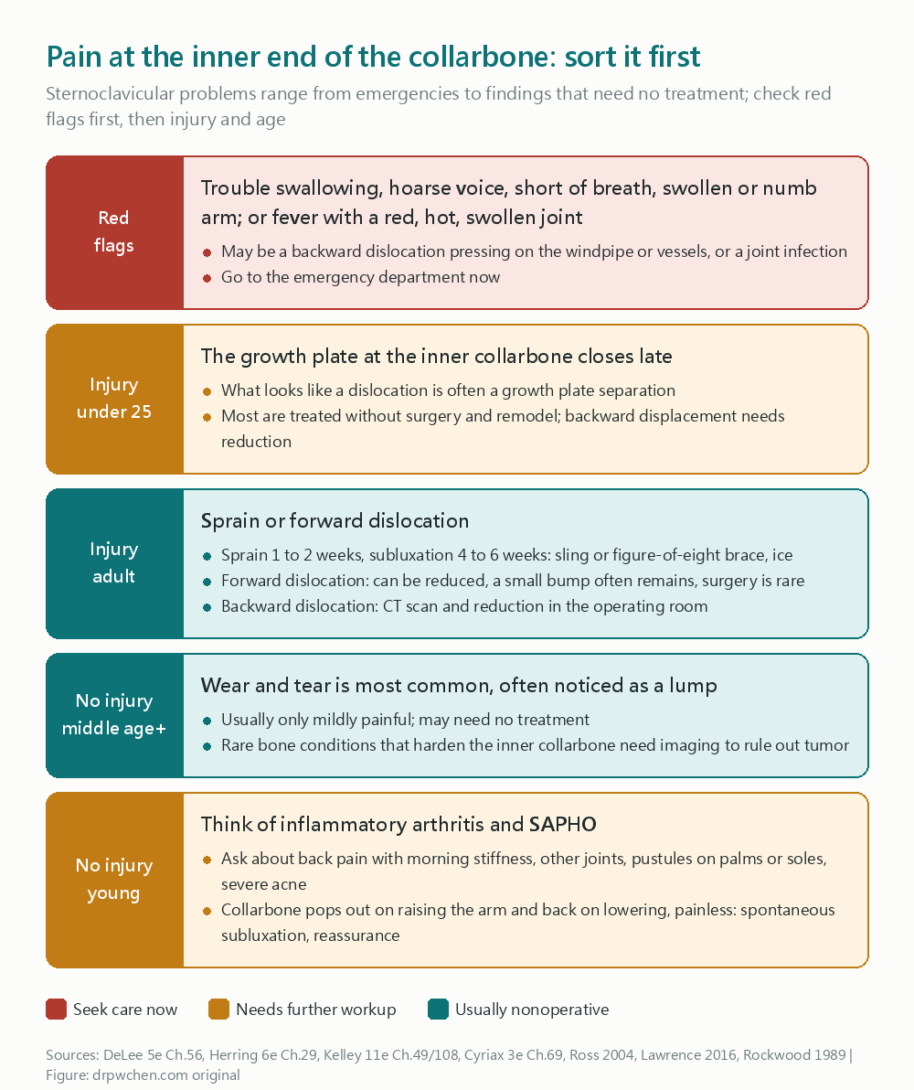
- Sprain: Ligaments are stretched, but the joint is still in place. A sling, ice, and pain relief usually get you back in about one to two weeks
- Subluxation: Ligaments are partially torn, and the joint is loose. Immobilization with a sling or a figure-of-eight brace for a few weeks, with recovery in about four to six weeks
- Dislocation: Ligaments are completely torn, and the medial clavicle pops out of the joint. This is further divided into anterior and posterior dislocations
Anterior dislocations are more common. You can feel a prominent bump on the inner collarbone. A doctor can help push it back in, but it’s often unstable and slips back out, usually leaving a slight bump in the end. The good news is this bump mostly just affects how it looks and rarely needs surgery.
Posterior dislocations are less common but more dangerous, as the collarbone is pushed backward into the windpipe, esophagus, and blood vessels. In this case, the inner collarbone actually looks sunken. This has to be confirmed with a CT scan, and the bone is pulled back into place in the operating room with a thoracic surgeon on standby.
Why do teenagers and young adults in their early twenties need special attention?
The growth plate at the medial clavicle is the last to close in the entire body. It often doesn’t finish growing until ages twenty-three to twenty-five.
So when a young person injures their medial clavicle, what looks like a dislocation is often actually a physeal separation: the bone slips right at the unfused growth plate. The upside of this injury is that the outer sleeve of the bone (the periosteum) is still attached and will slowly remodel on its own. So it is mostly treated conservatively with a sling or a figure-of-eight brace.
It’s only when it displaces backward and compresses the trachea or blood vessels behind it that the bone needs to be reduced in the operating room.
What if there was no injury, but the inner collarbone is swollen or constantly hurts?
This is actually more common than an injury. The reasons vary by age:
- Middle-aged and older, especially women: The most common cause is joint degeneration. Many people look in the mirror and notice a “bump” on their inner collarbone, but it doesn’t really hurt. CT scan studies show that most people over fifty have SC joint degeneration; it just mostly doesn’t cause symptoms
- Young adults: Consider inflammatory arthritis, like ankylosing spondylitis, psoriatic arthritis, or rheumatoid arthritis. The SC joint is frequently involved in these conditions
- Anterior chest pain with skin issues: If you have pustules on your palms or soles, severe acne, or hidradenitis suppurativa, along with pain around the front of your chest, collarbone, and breastbone, think about an inflammatory disease called SAPHO syndrome. This needs an evaluation by a rheumatologist
- The collarbone “pops out” when raising the arm, goes back in when lowered, and doesn’t hurt much: This is common in young people with lax ligaments and is called spontaneous anterior subluxation. It’s benign. Education and shoulder strengthening exercises are usually enough; studies show surgery tends to cause more problems here
- Rare bone diseases causing a swollen, painful, and sclerotic medial clavicle: Examples include condensing osteitis of the clavicle and aseptic necrosis of the medial clavicle (Friedrich disease). On imaging, these can look a lot like a tumor or infection. They require a doctor’s interpretation and sometimes a biopsy to confirm
Also, anterior chest pain might not be the SC joint at all. It could be a costal cartilage issue:
- If it hurts when you press on the joint between the ribs and the breastbone, but there’s no swelling, it’s more like costochondritis
- If there is swelling, and it’s mostly next to the second or third rib, it might be Tietze syndrome
Both of these are benign, but with any anterior chest pain, heart problems must be ruled out first.
Red, swollen, hot, painful inner collarbone: when to get it seen promptly
The SC joint can also get a bacterial infection. This is more likely to happen in:
- Patients with diabetes
- People with central venous catheters
- People who use intravenous drugs
- People who just had an infection somewhere else in their body
One thing to pay special attention to is that people with a joint infection don’t always have a fever. Many people have pain for a week or two before seeing a doctor. If your inner collarbone is red, swollen, hot, and getting more painful, please see a doctor as soon as possible. Doctors often use ultrasound to see how swollen the joint is and, if needed, draw joint fluid for culture.
What tests will be done?
- Ultrasound: Can show if there is fluid in the joint or if it’s dislocated, and allows comparison with the other side. If infection is suspected, fluid can also be drawn under ultrasound guidance
- X-ray: A standard chest X-ray isn’t great for seeing this joint; special angles are needed
- CT scan: The most important test when a dislocation or fracture is suspected after an injury, especially if a posterior dislocation is suspected
- MRI: Used in young people to distinguish between a physeal separation and a true dislocation, and also helps check for infection
When should you seek immediate medical attention?
- Difficulty swallowing, hoarse voice, shortness of breath
- Arm swelling, turning purple, numbness, or weakness
- The inner collarbone that used to bump out is now sunken in, and very painful
- Red, swollen, hot, and painful inner collarbone, or if accompanied by a fever
- Severe pain in a young person after an impact to the inner collarbone
—
Further reading: AC Joint Injuries, Shoulder Instability, Rotator Cuff Injuries, Shoulder Impingement
Below is the raw version of this note exactly as it appears in my vault, with only the internal links removed. The verification workflow and tools used are explained at the end.
Sternoclavicular Joint Disorders
Clinical Pearls
- The SC joint is the only true joint between the upper extremity and the torso. It offers almost no bony stability, relying heavily on the posterior capsule for anterior-posterior stability. The costoclavicular and interclavicular ligaments have little effect on AP translation
Spencer 2002,DeLee 5e Ch.56- Posterior dislocation is a medical emergency. The trachea, esophagus, and brachiocephalic vessels lie just behind it. Ask about dysphagia, hoarseness, dyspnea, and venous engorgement of the arm. CT (CTA if vascular injury is suspected). Reduction is performed under general anesthesia in the OR with a thoracic surgeon present
DeLee 5e Ch.56,Ingoe 2023- An “SC dislocation” in patients under 25 is mostly a medial clavicular physeal separation (the medial physis closes at 22–25 years). The periosteal sleeve is intact and will remodel extensively; management is primarily conservative
Herring 6e Ch.29,Magee Pathology 2e Ch.5- Non-traumatic SC pain: In middle-aged and older women, think OA first (CT shows nearly 90% of those >50 have degeneration, 0% in those <35). In young adults, look for SpA/RA, SAPHO, or spontaneous anterior subluxation. Fever + IVDU/diabetes/central venous catheter: septic arthritis until proven otherwise
Lawrence 2016,Ross 2004,Kelley 11e Ch.108- SC lesions can occasionally present with cervical signs (via the sternocleidomastoid [SCM]: turning to the asymptomatic side or resisted side-bending to the symptomatic side causes pain). Pain often refers to the shoulder, supraspinous fossa, and neck—neck rotation pain does not always equal facet joints
Ombregt Cyriax 3e Ch.69,Maigne 2e Ch.53
Abbreviations Glossary
| Abbreviation | Full term |
|---|---|
| SC / SCJ | sternoclavicular joint |
| OA | osteoarthritis |
| SpA | spondyloarthritis |
| RA | rheumatoid arthritis |
| SAPHO | synovitis, acne, pustulosis, hyperostosis, osteitis |
| IVDU | intravenous drug use |
| CTA | computed tomography angiography |
| RTS | return to sport |
Disease Background
Definitions
- Sternoclavicular joint = a saddle-type diarthrodial synovial joint between the medial clavicle, the clavicular notch of the manubrium, and the first costal cartilage. The only true joint between the upper extremity skeleton and the torso
Kelley 11e Ch.43,Martinoli Ch.6- The clavicular articular surface is much larger than the shallow sternal socket. Only the lower half of the medial clavicle contacts the sternum → intrinsically bony unstable
Kahn 1e Ch.35,ElMiedany 1e Ch.12 - In about 2.5% of people, the medial clavicle also forms a joint with the superior border of the first rib
DeLee 5e Ch.56
- The clavicular articular surface is much larger than the shallow sternal socket. Only the lower half of the medial clavicle contacts the sternum → intrinsically bony unstable
- This note covers two broad categories (single joint, distinct etiological spectrums):
- Trauma: sprain, anterior/posterior dislocation, medial clavicular physeal separation, chronic instability
- Non-trauma: SC OA, condensing osteitis, Friedrich disease, SAPHO, septic arthritis, RA/SpA, spontaneous atraumatic anterior subluxation
Anatomy and Stabilizing Structures
- Stability relies entirely on soft tissues: capsular lig. (anterior, superior, posterior), costoclavicular (rhomboid) lig., interclavicular lig., intra-articular disk lig.
DeLee 5e Ch.56- Intra-articular disk: Originates from the synchondrosis of the first rib and sternum, inserts on the posterosuperior margin of the medial clavicle, dividing the joint into two cavities → prevents the clavicle from medial displacement
DeLee 5e Ch.56,Magee Pathology 2e Ch.5 - Costoclavicular lig.: Anterior fasciculus limits lateral displacement and excessive upward rotation; posterior fasciculus limits medial displacement and excessive downward rotation
Magee Pathology 2e Ch.5 - Interclavicular lig.: Assists the capsular lig. in maintaining shoulder poise; taut when the arm is resting at the side, relaxed upon elevation
Magee Pathology 2e Ch.5 - Capsular lig.: The strongest ligament; prevents the medial clavicle from tilting upward when a downward force is applied to the lateral shoulder
DeLee 5e Ch.56
- Intra-articular disk: Originates from the synchondrosis of the first rib and sternum, inserts on the posterosuperior margin of the medial clavicle, dividing the joint into two cavities → prevents the clavicle from medial displacement
- Biomechanics (cadaver sectioning studies)
Spencer 2002- The posterior capsule is the most important restraint against both anterior and posterior translation; the anterior capsule primarily restrains anterior translation.
- Sectioning the costoclavicular or interclavicular ligaments has minimal effect on AP translation.
- Cited in textbooks: the force required to produce a posterior dislocation is about 1.5 times that required for an anterior dislocation
Magee Pathology 2e Ch.5,DeLee 5e Ch.56
- Range of motion: During elevation, the clavicle elevates about 35°, moves anteriorly/posteriorly about 35°, and rotates 45–50° along its long axis
DeLee 5e Ch.56,Magee Pathology 2e Ch.5- Almost all upper extremity movements involve the SC joint → clinically, SC lesions will implicate shoulder and shoulder girdle movements
Magee Pathology 2e Ch.5
- Almost all upper extremity movements involve the SC joint → clinically, SC lesions will implicate shoulder and shoulder girdle movements
- Posterior structures at risk: trachea, esophagus, brachiocephalic vein/trunk, subclavian artery, common carotid artery, internal jugular vein, phrenic and vagus nerves
DeLee 5e Ch.56,Herring 6e Ch.29 - Medial clavicular physis: The clavicle is the first bone in the body to ossify and the last to close.
- The medial epiphysis does not appear until ages 18–20, and fuses with the diaphysis at ages 23–25; Kahn cites closure as late as 31 years
Magee Pathology 2e Ch.5,Kahn 1e Ch.35 - Herring notes closure at 22–25 years
Herring 6e Ch.29
- The medial epiphysis does not appear until ages 18–20, and fuses with the diaphysis at ages 23–25; Kahn cites closure as late as 31 years
Epidemiology
- Traumatic SC injuries are rare
- DeLee: accounts for at most 3% of shoulder girdle injuries
DeLee 5e Ch.56; Magee: about 3% of shoulder girdle fractures and dislocationsMagee Pathology 2e Ch.5 - Single Level I trauma center with 92 cases (1992–2011): accounted for 0.9% of shoulder girdle injuries, 1.1% of all dislocations; average age 39, primary mechanism was a fall, 16% were physeal injuries
Boesmueller 2016
- DeLee: accounts for at most 3% of shoulder girdle injuries
- Anterior dislocations are more common than posterior, though reported ratios vary widely.
- Magee: Anterior is 2–3 times more common than posterior
Magee Pathology 2e Ch.5 - Cyriax: Anterior can be up to 20 times more common than posterior
Ombregt Cyriax 3e Ch.69
- Magee: Anterior is 2–3 times more common than posterior
- Adolescents: medial physeal fractures account for 1–6% of clavicle injuries in skeletally immature patients
DeLee 5e Ch.133 - Non-traumatic
- SC OA is extremely common on CT (all incidental findings on CTs taken for other reasons): 59% of 232 patients had OA signs on at least one side; 0% in those under 35, 89.6% in those over 50, 100% in those over 61
Lawrence 2016- Maigne: At age 60, SC degeneration is “almost invariably present” and appears later than AC degeneration
Maigne 2e Ch.53
- Maigne: At age 60, SC degeneration is “almost invariably present” and appears later than AC degeneration
- Septic arthritis: accounts for about 1% of septic arthritis in the general population, but 17% in IVDU
Ross 2004; in joint distribution data cited by Kelley, the SC joint accounts for 5%Kelley 11e Ch.116 - SAPHO: estimated prevalence of roughly 1 in 10,000, likely underestimated (in those with mild or no skin manifestations)
Kelley 11e Ch.108
- SC OA is extremely common on CT (all incidental findings on CTs taken for other reasons): 59% of 232 patients had OA signs on at least one side; 0% in those under 35, 89.6% in those over 50, 100% in those over 61
Mechanism of Injury
Trauma
- Mostly high-energy: MVAs are the most common, followed by sports injuries
DeLee 5e Ch.56,Magee Pathology 2e Ch.5 - Indirect force is the most common: force transmitted from the lateral shoulder to the SC joint
DeLee 5e Ch.56,Kahn 1e Ch.35- Lying on the side and being compressed, with the shoulder rolled backward → anterior dislocation
- Shoulder rolled forward (e.g., in a football pile-up) → posterior dislocation
- Direct force to the anterior medial clavicle (steering wheel, getting kicked, hitting a goalpost) → almost always results in a posterior dislocation
DeLee 5e Ch.56,Kahn 1e Ch.35,Magee Pathology 2e Ch.5 - Adolescent physeal separation: the diaphysis protrudes through the anterior or posterior periosteal sleeve, while the epiphysis remains attached to the SC/costoclavicular ligaments
Herring 6e Ch.29,DeLee 5e Ch.133
Non-Trauma
- Spontaneous atraumatic anterior subluxation: In individuals with ligamentous laxity, the medial clavicle slips out anteriorly when raising the arm overhead and spontaneously reduces when lowered, mostly painless
DeLee 5e Ch.56,Rockwood 1989 - OA: More common on the dominant side of postmenopausal women
Ombregt Cyriax 3e Ch.69,Martinoli Ch.6- Spinal accessory nerve injury from neck surgery → drooping and forward-protracted shoulder, increased SC loading, is considered a predisposing factor
Martinoli Ch.6
- Spinal accessory nerve injury from neck surgery → drooping and forward-protracted shoulder, increased SC loading, is considered a predisposing factor
- Septic: Bacteria may enter the joint directly from near the subclavian vein valves or from injections between the two heads of the SCM
Ross 2004 - SAPHO: Etiology unknown; hypotheses include chronic Propionibacterium acnes (P. acnes) infection, Th17/Treg imbalance; unrelated to HLA-B27
Kelley 11e Ch.108
Complications
- Anterior dislocation: The most common is a prominent bump (the medial clavicle remains prominent after conservative treatment), followed by recurrent instability and late-stage degeneration
DeLee 5e Ch.56,Magee Pathology 2e Ch.5 - Posterior dislocation (acute): pneumothorax, laceration or occlusion of great vessels, esophageal rupture, brachial plexus compression, recurrent laryngeal nerve injury causing voice changes
DeLee 5e Ch.56- Late: tracheoesophageal fistula, stridor, dysphagia
DeLee 5e Ch.56 - Glass systematic review: among 80 posterior dislocations, 30% had symptoms of mediastinal compression (cited in DeLee)
DeLee 5e Ch.56,Glass 2011 - Magee: severe complications can be as high as 25%, with about 3 reported deaths in the literature
Magee Pathology 2e Ch.5
- Late: tracheoesophageal fistula, stridor, dysphagia
- Surgical complications: infection, loss of reduction, post-traumatic arthritis, neurovascular injury; K-wire migration has caused deaths and near-deaths → K-wires are strictly contraindicated for the SC joint
DeLee 5e Ch.56,DeLee 5e Ch.133 - Septic SC: osteomyelitis 55%, chest wall abscess/phlegmon 25%, mediastinitis 13%
Ross 2004
Prognosis
- Traumatic prognosis is generally good: of 92 cases, 49% completed long-term follow-up (median 11.3 years), with a mean American Shoulder and Elbow Surgeons (ASES) score of 96 out of 100
Boesmueller 2016 - Glass 2011 systematic review (24 case series, 251 cases; cited in DeLee)
DeLee 5e Ch.56,Glass 2011- Anterior dislocation good–excellent: nonoperative 69%, closed reduction 85%, open reduction after failed closed 80%, direct open 75%
- Posterior dislocation good–excellent: closed reduction 100%, open reduction after failed closed 88%, direct open 91%
- Attempting closed reduction first does not worsen the outcomes of subsequent open reduction; acute is better than chronic
- After closed reduction of posterior dislocation: 74% of 31 cases returned fully to their original sport at an average of 3.1 months
Damschen 2022 - Physeal separation: intact periosteal sleeve, strong remodeling potential, mostly no long-term sequelae
Herring 6e Ch.29,DeLee 5e Ch.133 - Spontaneous anterior subluxation: benign natural history; among 37 cases, all 29 in the conservative group were excellent at an average 8-year follow-up, while the 8 who had surgery suffered from scarring, persistent instability, and pain
Rockwood 1989
Clinical Evaluation
Diagnostic Criteria
- No formal diagnostic criteria; clinical diagnosis + imaging
- Trauma: history + local point tenderness/deformity + imaging (primarily CT)
DeLee 5e Ch.56 - Non-trauma: Defined by the respective etiology, see “Differential Diagnosis” below
- Trauma: history + local point tenderness/deformity + imaging (primarily CT)
Evaluation Tools
History
- Mechanism of trauma, direction of force (see “Mechanism of Injury” above)
DeLee 5e Ch.56 - Posterior dislocation red flags: dyspnea, choking sensation, dysphagia, hoarseness, arm numbness or weakness, arm swelling and cyanosis
Kahn 1e Ch.35,DeLee 5e Ch.133- Mediastinal compression can be present immediately upon injury, or it can appear delayed
Groh 2011
- Mediastinal compression can be present immediately upon injury, or it can appear delayed
- Age <25 → consider physeal separation
Kahn 1e Ch.35,Groh 2011 - Non-trauma: fever, IVDU, DM, central venous catheter, distant infection, RA
Ross 2004,Ombregt Cyriax 3e Ch.69- Skin: palmoplantar pustulosis, severe acne, hidradenitis suppurativa, psoriasis (can precede or follow bone/joint involvement by years) → SAPHO
Kelley 11e Ch.108 - Inflammatory back pain, morning stiffness, other joints → SpA/RA
Ombregt Cyriax 3e Ch.69 - Medial clavicle “pops out” on elevation, reduces on lowering, painless → spontaneous subluxation
DeLee 5e Ch.56
- Skin: palmoplantar pustulosis, severe acne, hidradenitis suppurativa, psoriasis (can precede or follow bone/joint involvement by years) → SAPHO
- Location of pain: SC pain is often not at the SC joint itself; it refers to the shoulder, supraspinous fossa, and neck
Maigne 2e Ch.53- Posterior sternoclavicular syndrome: spontaneous posterior root-of-neck pain in middle-aged patients, mimics a cervical spine issue but cervical exam is negative
Ombregt Cyriax 3e Ch.69
- Posterior sternoclavicular syndrome: spontaneous posterior root-of-neck pain in middle-aged patients, mimics a cervical spine issue but cervical exam is negative
Physical Examination
- Posture: Patient supports the affected arm with the contralateral hand, head tilted toward the affected side (to reduce SC loading)
DeLee 5e Ch.56,Herring 6e Ch.29- Severe cases cannot lie flat
DeLee 5e Ch.56
- Severe cases cannot lie flat
- Inspection
- Anterior dislocation: medial clavicle prominent and palpable
Kahn 1e Ch.35 - Posterior dislocation: medial clavicle sunken, indistinct; swelling may mask the difference
Kahn 1e Ch.35,Magee Pathology 2e Ch.5 - Appearance and direction are unreliable: swelling will mask it; posterior dislocation absolutely requires imaging to confirm
DeLee 5e Ch.133,Kahn 1e Ch.35
- Anterior dislocation: medial clavicle prominent and palpable
- PE findings by grade
DeLee 5e Ch.56- Type I: mild pain on movement, mild joint swelling, and point tenderness
- Type II: same as above + manual stress testing reveals subluxation
- Type III: arm adducted, head tilted to the affected side, severe pain with any movement
- Must do: Bilateral upper extremity pulses, sensation, motor function, jugular venous distention
Kahn 1e Ch.35,Magee Pathology 2e Ch.5 - Cyriax three-tier signs (all SC lesions)
Ombregt Cyriax 3e Ch.69- Neck: traction via SCM → active/resisted rotation to the asymptomatic side, resisted side-bending to the symptomatic side causes pain
- Shoulder girdle: active and passive shoulder girdle elevation causes pain
- Shoulder: elevation is most painful; passive horizontal adduction is the most painful provocative movement
- Other: lateral compression of the shoulder causes pain, pain raising arm over 90°; a torn disk can have meniscus-like mechanical symptoms
Kahn 1e Ch.35 - Non-trauma clues: Inflammatory arthritis presents with local swelling due to synovial thickening
Ombregt Cyriax 3e Ch.69; septic has a local red, hot mass (abscess) in about 20% of casesOmbregt Cyriax 3e Ch.69
Radiographs
- Standard AP chest: too much overlap, not suitable for viewing the SC joint, but can check for clavicular asymmetry and pneumothorax
DeLee 5e Ch.56 - Special views: the Heinig view (supine, beam tangential to the joint) and the Hobbs view (patient leaning forward over the cassette, beam from above) are better than AP but still limited
DeLee 5e Ch.56 - Serendipity view (Rockwood): X-ray tube tilted 40° cephalad from the vertical, aimed at the manubrium
DeLee 5e Ch.56- Anterior dislocation: the affected medial clavicle is projected above the horizontal line of the normal side; posterior dislocation is projected below it
DeLee 5e Ch.56,Kahn 1e Ch.35 - Tube distance is 45 inches for children, 60 inches for adults
DeLee 5e Ch.56
- Anterior dislocation: the affected medial clavicle is projected above the horizontal line of the normal side; posterior dislocation is projected below it
- Non-trauma: OA shows osteophytes, bone cysts, hyperostosis, joint space narrowing; most prominent at the inferior margin of the clavicular head
Ombregt Cyriax 3e Ch.69
Computed Tomography
- The imaging modality of choice for SC trauma: can differentiate fractures from dislocations, and detect mild subluxations
DeLee 5e Ch.56,Groh 2011- Posterior dislocation: get a CTA first to check the distance between the clavicle and the innominate vein, and check for hemorrhage
Ingoe 2023 - Angiography is required if vascular or mediastinal compression is suspected
Kahn 1e Ch.35
- Posterior dislocation: get a CTA first to check the distance between the clavicle and the innominate vein, and check for hemorrhage
- Non-trauma: CT is better than X-rays for visualizing osteitis/hyperostosis in flat bones (sternum, clavicle)
Kelley 11e Ch.108- Beware of incidental OA: nearly 90% of those over 50 have SC degeneration on CT; keep this in mind when evaluating a symptomatic SC joint
Lawrence 2016 - RA’s CT appearance in the SC joint is difficult to distinguish from age-related degeneration
Celikyay 2013 - Degeneration more commonly shows subchondral cysts (on CT)
Johnson 2010
- Beware of incidental OA: nearly 90% of those over 50 have SC degeneration on CT; keep this in mind when evaluating a symptomatic SC joint
Magnetic Resonance Imaging
- Used in patients under 23 to distinguish between physeal injury and true dislocation
Groh 2011 - Septic: fluid signal marrow replacement is significantly associated with infection
Johnson 2010; MRI can also show soft tissue abscessesKelley 11e Ch.116 - Fixation of physeal fractures is recommended with sutures rather than metal to avoid interfering with future MRIs
Herring 6e Ch.29
Ultrasonography
- Can diagnose anterior/posterior dislocations and subluxations, dynamic scanning and comparison with the asymptomatic side
Jacobson 3e Ch.3- If posterior dislocation is suspected, always follow up with a CT to check vessels
Jacobson 3e Ch.3 - Can be used intraoperatively to confirm posterior reduction
Martinoli Ch.6
- If posterior dislocation is suspected, always follow up with a CT to check vessels
- Infection vs Degeneration (Johnson 2010; a retrospective study of 19 pathologically confirmed cases)
Johnson 2010- Infection: joint capsule distention averaged 14 mm (10–20 mm), with 60% overlying both the clavicle and sternum
- Degeneration: averaged 5 mm (3–8 mm), overlying only the clavicular side
- Other factors significantly associated with infection: MRI marrow fluid signal, elevated Westergren ESR, and fever
- ⚠️ Hyperemia was not a statistically significant differentiator in the original paper; ElMiedany lists “color Doppler (CD) hyperemia” alongside >10 mm distention as a septic finding
ElMiedany 1e Ch.12—hyperemia is a common concomitant finding, but not a validated differentiator confirmed by Johnson. - If infection is suspected, perform ultrasound-guided aspiration; degenerative SC capsular thickening in older women is often palpated as a “mass”
Jacobson 3e Ch.3
- RA: about 40% have SC synovitis; ultrasound detection rate is roughly twice that of physical examination
ElMiedany 1e Ch.12 - OA: joint space narrowing, osteophytes, para-articular cysts
Martinoli Ch.6
Laboratory Tests
- Septic: elevated ESR; only 65% have fever, 62% have bacteremia
Ross 2004,Johnson 2010 - SAPHO: ESR/CRP are only moderately elevated, unreliable
Kelley 11e Ch.108 - Joint fluid: aspiration for culture if infection is suspected (see ultrasound section above)
Jacobson 3e Ch.3
Classification Systems
Trauma: Severity (Allman/Rockwood 3-Tier Classification)
- Grading drives management; treatment for each grade is outlined in the Management Algorithm below.
| Grade | Ligaments | Clinical | Source |
|---|---|---|---|
| I (mild sprain) | SC lig. partially torn, costoclavicular lig. intact | Mild pain, no instability | DeLee 5e Ch.56, Kahn 1e Ch.35 |
| II (subluxation) | SC lig. completely torn; costoclavicular lig. intact or partially torn | Mild deformity, palpable subluxation | DeLee 5e Ch.56, Magee Pathology 2e Ch.5 |
| III (dislocation) | SC and costoclavicular lig. both torn | Obvious swelling and deformity, severe pain | DeLee 5e Ch.56, Kahn 1e Ch.35 |
- Note: DeLee originally wrote “CC ligament”, which in the context of the SC joint means costoclavicular, not the coracoclavicular ligament of the AC joint
DeLee 5e Ch.56 - Costoclavicular lig. in Type II: Kahn notes it as intact, Magee notes it as a 2nd-degree sprain (partially torn)
Kahn 1e Ch.35,Magee Pathology 2e Ch.5
Direction, Etiology, and Timing
- Direction: anterior (common) / posterior (dangerous)
DeLee 5e Ch.56 - Etiology and timing: sprain/subluxation, acute dislocation, recurrent, unreduced, atraumatic
DeLee 5e Ch.56,Kahn 1e Ch.35 - Age: <25 years, first rule out physeal separation (pseudodislocation)
Herring 6e Ch.29,Groh 2011
Differential Diagnosis
| Category | Diagnosis | Key Differentiators | Source |
|---|---|---|---|
| Trauma | Medial clavicular physeal separation | Age <25; epiphysis stays in place, diaphysis displaces; MRI/CT to differentiate | Herring 6e Ch.29, Groh 2011 |
| Trauma | Medial clavicle fracture, sternal fracture, rib fracture | CT differentiates fracture from dislocation | DeLee 5e Ch.56, Kahn 1e Ch.35 |
| Degeneration | SC OA | Postmenopausal women, primarily presents as visual thickening, mild pain; extremely common incidental finding on CT | Ombregt Cyriax 3e Ch.69, Lawrence 2016 |
| Degeneration | Sternoclavicular hyperostosis | Young onset, intermittent painful, swollen, red sternum and SC joint, progressive ossification to ankylosis; can be part of SAPHO | Ombregt Cyriax 3e Ch.69 |
| Inflammation | SAPHO | Anterior chest wall pain is the most typical presentation; skin findings; bone scan bull’s head sign (increased uptake in the manubrium and both SC joints); about 1/3 have spinal involvement | Kelley 11e Ch.108 |
| Inflammation | RA | SC has synovium but little movement, rarely symptomatic; frank SC inflammation in an RA patient must rule out infection first | Kelley 11e Ch.76 |
| Inflammation | SpA (ankylosing spondylitis [AS], psoriatic arthritis [PsA]) | SC frequently involved, might be the first joint; can progressively ankylose | Kelley 11e Ch.43, Ombregt Cyriax 3e Ch.69 |
| Inflammation | Crystal (gout, calcium pyrophosphate deposition [CPPD]) | CPPD can deposit in the SC joint | DeLee 5e Ch.56, ElMiedany 1e Ch.7 |
| Infection | Septic arthritis | IVDU, DM, central venous catheter, distant infection; fever in only 65%; US distention ≥10 mm crossing both bones | Ross 2004, Johnson 2010 |
| Bone Disease | Condensing osteitis of the clavicle | Middle-aged women, medial 1/3 clavicle tender swelling, osteosclerosis; imaging easily confused with tumor | Kelley 11e Ch.49, Davis 2022 |
| Bone Disease | Friedrich disease | Aseptic necrosis of the sternal end of the clavicle; can spontaneously resolve, easily misdiagnosed as osteomyelitis or tumor | Kelley 11e Ch.49, Christensen 1987 |
| Instability | Spontaneous atraumatic anterior subluxation | Ligamentous laxity, slips out when raising arm, mostly painless | Rockwood 1989, Kahn 1e Ch.35 |
| Chest Wall | Tietze syndrome | Upper costal cartilage (mostly 2nd-3rd) with swelling, unilateral, single site, young people; can also involve SC | Frontera 4e Ch.117, Kelley 11e Ch.49 |
| Chest Wall | Costochondritis/costosternal syndrome | Costosternal joint tenderness without swelling, multiple sites, mostly women over 40 | Frontera 4e Ch.117, Kelley 11e Ch.43 |
| Referred Pain | Cervical spine origin (facet, radiculopathy) | Cervical exam can reproduce symptoms, SC locally non-tender; however, SC lesions themselves can also cause cervical signs | Ombregt Cyriax 3e Ch.69, Maigne 2e Ch.53 |
| Other | AC lesions, apical lung lesions, first rib fracture | Cyriax groups these together under “clavipectoral pain” shoulder girdle lesions | Ombregt Cyriax 3e Ch.69 |
Non-Trauma Etiology Details
- SC OA
Ombregt Cyriax 3e Ch.69,Martinoli Ch.6,Lawrence 2016- Chief complaint is often visual thickening, with mild pain; shoulder girdle mobility restriction leads to limited elevation.
- 40–60 year old women on the dominant side
Martinoli Ch.6 - Painful only during “overuse”: overusing an already degenerated joint will cause a sprain
Ombregt Cyriax 3e Ch.69
- Condensing osteitis of the clavicle
- Rare benign idiopathic lesion, located in the medial 1/3 of the clavicle; a more precise name is aseptic enlarging osteosclerosis; middle-aged women, tender swelling of the medial 1/3 of the clavicle
Kelley 11e Ch.49 - Imaging overlaps with benign and malignant bone lesions, biopsy is often required
Davis 2022 - Also reported in children (7 cases averaging 11.5 years old, biopsy confirmed)
Andreacchio 2016 - European pediatric cancer registries: medial clavicle malignancies are extremely rare; in the same paper’s literature review, only 2 of 89 biopsies grew an organism → clinically typical cases can be followed with serial imaging; biopsy only if in doubt
Clement 2011
- Rare benign idiopathic lesion, located in the medial 1/3 of the clavicle; a more precise name is aseptic enlarging osteosclerosis; middle-aged women, tender swelling of the medial 1/3 of the clavicle
- Friedrich disease
- Painful aseptic necrosis of the sternal end of the clavicle
Kelley 11e Ch.49 - Often resolves spontaneously; a correct diagnosis avoids unnecessary treatment; can lead to early SC degeneration
Christensen 1987,Macule 1990
- Painful aseptic necrosis of the sternal end of the clavicle
- SAPHO
Kelley 11e Ch.108- Early histology of bone lesions resembles osteomyelitis, followed by chronic inflammation, and late-stage fibrosis and sclerosis.
- Anterior chest wall X-ray/CT: homogeneous fibrous osteosclerosis, periosteal reaction, cortical thickening; erosive arthritis typically at the SC, upper costosternal, and manubriosternal joints.
- High suspicion: multifocal recurrent sterile osteomyelitis; arthritis combined with palmoplantar pustulosis / pustular psoriasis / severe acne.
- Septic SC (review of 180 cases)
Ross 2004- Mean age 45, 73% male; chest pain 78%, shoulder pain 24%; median of 14 days of symptoms before seeking care.
- Risk factors: IVDU 21%, distant infection 15%, DM 13%, trauma 12%, infected central venous catheter 9%; 23% had no risk factors.
- Pathogen: S. aureus 49%
- Cyriax adds: immunocompromised elderly, RA, DM, drug addiction; Brucella has also been reported
Ombregt Cyriax 3e Ch.69
Red Flags and Referrals
- Immediate referral to ER/Trauma Center (Advanced Trauma Life Support [ATLS] ABCs)
Kahn 1e Ch.35- Respiratory distress, dysphagia, hoarseness
- Upper extremity venous engorgement, diminished pulses, cyanosis, paresthesias
- Hemodynamic instability
- Suspected posterior dislocation or posteriorly displaced physeal fracture
- Fever + red, swollen, hot, painful SC joint → same-day aspiration/imaging to rule out septic arthritis
Kahn 1e Ch.35,Jacobson 3e Ch.3- Confirmed septic: CT or MRI to rule out chest wall phlegmon, retrosternal abscess, mediastinitis; if present, requires thoracic surgery en-bloc resection
Ross 2004
- Confirmed septic: CT or MRI to rule out chest wall phlegmon, retrosternal abscess, mediastinitis; if present, requires thoracic surgery en-bloc resection
- Orthopedic referral: Type III dislocation, suspected physeal fracture
Kahn 1e Ch.35 - Rheumatology referral: multifocal osteitis, skin manifestations, inflammatory back pain, polyarthritis
- Unexplained sclerotic lesion of the medial clavicle → bone tumor/infection workup, biopsy if necessary
Clement 2011,Davis 2022
Clinical Management
Management Algorithm
- There are no society-level clinical practice guidelines (CPGs) for this condition (full text search in
_Guidelines/yields no SC topics; literature consists entirely of case series and Level IV systematic reviews)Obremskey 2022,Martetschläger 2014 - Axis driving management: Trauma vs Non-trauma → Direction (prioritize ruling out posterior) → Age (physis) → Severity
Algorithm
- Step one: Presence of mediastinal or neurovascular symptoms (dysphagia, hoarseness, dyspnea, arm venous engorgement or weak pulses) → Yes = emergency, ATLS + CTA + thoracic/vascular surgery
Kahn 1e Ch.35,Ingoe 2023- Trauma
- Type I: Ice, analgesics, sling, progressive ROM as tolerated; ~1–2 weeks to recover
DeLee 5e Ch.56,Kahn 1e Ch.35- Type II: Ice, analgesics, clavicle strap/sling and swathe/figure-of-eight for 1 week, then a sling for another 4–6 weeks; if subluxed, can gently push both shoulders posteromedially to reduce in clinic
DeLee 5e Ch.56- Type III anterior dislocation: Closed reduction under sedation or general anesthesia → figure-of-eight for at least 6 weeks, protect for another 2 weeks before strenuous activity; often unstable and will slip out again, but rarely needs surgery
DeLee 5e Ch.56- Type III posterior dislocation: CT/CTA → closed reduction in the OR under general anesthesia with a thoracic surgeon present → figure-of-eight for 4–6 weeks; if closed reduction fails or mediastinum is compressed → open reduction
DeLee 5e Ch.56,Ingoe 2023,Sernandez 2019
- High success rate for closed reduction within 48 hours; can still be attempted within 10 days post-injury
Ingoe 2023- <25 years: Confirm if it’s a physeal separation first (MRI/CT) → see branches below
Groh 2011
- Anterior displacement or posterior displacement without mediastinal compression: sling or figure-of-eight for symptomatic treatment; if cosmesis is very poor, can try closed reduction, if it pops out just accept it and wait for remodeling
Herring 6e Ch.29- Posterior displacement + airway/esophageal/neurovascular compression: emergency OR closed reduction; if it fails, proceed to open reduction and ligament repair, ideally with thoracic surgery
Herring 6e Ch.29- Late-presenting posterior physeal fracture is an absolute contraindication for closed reduction (may have adhered to mediastinal vessels)
Herring 6e Ch.29- Chronic instability that remains symptomatic after conservative management → then consider reconstruction (figure-of-eight tendon graft)
DeLee 5e Ch.56,Martetschläger 2014- Non-trauma
- Rule out septic first (fever, risk factors, US distention ≥10 mm crossing both bones) → aspirate for culture, antibiotics, surgery if necessary
Johnson 2010,Ross 2004- Spontaneous anterior subluxation: education and reassurance, shoulder strengthening, no surgery, avoid intentionally popping it out
Rockwood 1989,Kahn 1e Ch.35- OA: mostly requires no treatment; painful overuse → intra-articular steroid; conservative failure, chronic pain → resection arthroplasty (preserving the costoclavicular lig.)
Ombregt Cyriax 3e Ch.69,DeLee 5e Ch.56- Inflammatory (RA/SpA/SAPHO): treat the underlying disease; SAPHO first-line is NSAIDs
Kelley 11e Ch.108
Treatment Goals
- Pain control, reduction and immobilization as needed, manage concomitant injuries, educate on protection and prevention of re-injury
Magee Pathology 2e Ch.5 - Be mindful of compensatory movement patterns of the entire shoulder complex following an SC injury
Magee Pathology 2e Ch.5
Conservative Management
Traumatic Sprain / Subluxation
- Stable Type I–II: Immobilize for 3–4 days (based on pain), then progressively return to normal use; electrotherapy, ice/heat can be used for pain relief
Magee Pathology 2e Ch.5 - Return to activity: Type I ~1–2 weeks, Type II ~4–6 weeks
Kahn 1e Ch.35 - Physeal fractures typically heal in 3–4 weeks; requires pain-free full ROM and strength recovery before returning to sports
Kahn 1e Ch.35 - Type III: Return to sports is decided by a multidisciplinary team
Kahn 1e Ch.35
Closed Reduction Techniques
- Anterior dislocation: Supine, place a 3–4 inch pad between the scapulae to retract them → push the medial clavicle posteriorly
DeLee 5e Ch.56 - Posterior dislocation (abduction–traction): Supine with interscapular pad, abduct the arm and apply lateral traction while slowly extending; if reduction is difficult, use fingers or a sterile towel clip to pull the clavicle anterolaterally
DeLee 5e Ch.56 - Structures potentially injured in posterior dislocation: trachea, brachiocephalic vein/trunk, subclavian artery, common carotid artery
DeLee 5e Ch.56 - Both closed and open reduction can result in massive hemorrhage; recommended that the entire team rehearses the bleeding protocol prior to reduction, and high-risk patients should have venous lines placed in advance
Ingoe 2023
Spontaneous Anterior Subluxation
- Gentle shoulder strengthening, analgesics, heat; avoid intentionally popping it out
Kahn 1e Ch.35 - Conservative management yields no long-term activity restrictions; surgical outcomes are poor (scarring, persistent instability, requiring multiple resections, even thoracic outlet syndrome)
Rockwood 1989
Injections
- Intra-articular triamcinolone: can be used for acute OA overuse, RA, sprains
Ombregt Cyriax 3e Ch.69- Technique: Supine, palpate the gap between the medial clavicle and sternum, advance a 2.5 cm needle about 1 cm, inject 1 mL; high resistance indicates being within the disk, withdraw slowly while applying pressure until flow is smooth
Ombregt Cyriax 3e Ch.69 - Posterior sternoclavicular syndrome (posterior ligament lesion; see History above): infiltrate 20 mg into the posterior ligament from a superior approach or trans-articularly, must avoid puncturing posterior viscera; re-evaluate at 2 weeks, usually requires 2–3 sessions
Ombregt Cyriax 3e Ch.69
- Technique: Supine, palpate the gap between the medial clavicle and sternum, advance a 2.5 cm needle about 1 cm, inject 1 mL; high resistance indicates being within the disk, withdraw slowly while applying pressure until flow is smooth
- Ultrasound-guided: in-plane, lateral to medial approach, avoiding deep joint structures
Jacobson 3e Ch.9- Image guidance accuracy for upper extremity joints (including SC) is superior to blind palpation
Patel 2022
- Image guidance accuracy for upper extremity joints (including SC) is superior to blind palpation
- ⚠️ No controlled trials on the efficacy of SC injections were found for this note; injections are for symptom control, with an evidence level of expert opinion and case series.
Pharmacotherapy
- SAPHO: NSAIDs are first-line but often insufficient → steroids, bisphosphonates (especially pamidronate, mostly complete or partial response), TNF inhibitors (less effective than in other SpA, skin symptoms may flare); if failed, can use IL-1 or IL-17/IL-23 inhibitors
Kelley 11e Ch.108- Antibiotics (azithromycin) have a transient effect that fades after discontinuation, mechanism unknown
Kelley 11e Ch.108
- Antibiotics (azithromycin) have a transient effect that fades after discontinuation, mechanism unknown
- Septic: Antibiotics based on culture; empiric therapy must consider MRSA coverage
Ross 2004 - Condensing osteitis (pediatric series): IV + oral antibiotics post-biopsy, avoiding aggressive surgery
Andreacchio 2016- ⚠️ Single 7-case series, no control group; impossible to determine if treatment was effective or if it resolved naturally.
Surgical Management
Indications
- Trauma: chronic symptomatic instability, failed or unstable closed reduction of posterior dislocation, mediastinal compression
DeLee 5e Ch.56,Martetschläger 2014- Surgical risks often outweigh the benefits: infection, recurrence, poor cosmesis, neurovascular injury, implant migration → considered only if chronic symptomatic instability persists after a period of conservative management
DeLee 5e Ch.56
- Surgical risks often outweigh the benefits: infection, recurrence, poor cosmesis, neurovascular injury, implant migration → considered only if chronic symptomatic instability persists after a period of conservative management
- OA: persistent pain and failed conservative management
Martetschläger 2014 - Septic: chest wall phlegmon, retrosternal abscess, mediastinitis → en-bloc resection, can be supplemented with a pectoralis major flap
Ross 2004
Figure-of-Eight Tendon Graft Reconstruction
- Current procedure of choice for SC instability: hamstring or palmaris autograft, or semitendinosus allograft, woven in a figure-of-eight fashion through drill holes in the clavicle and manubrium
DeLee 5e Ch.56 - Cadaveric biomechanics: figure-of-eight semitendinosus failure load is ~230–240 N, superior to intramedullary ligament and subclavius tendon reconstructions (~50–85 N)
Spencer 2004 - Systematic review: figure-of-eight tendon reconstruction is recommended for chronic instability; simple capsular repair is sufficient for acute cases requiring open reduction
Thut 2011 - Reconstruction for chronic instability (12 studies, 164 patients): patient-reported outcome scores all significantly improved, overall complications low; complication rate bicortical 9% vs unicortical 15% (no significant diff), unicortical trended higher for recurrent instability (10% vs 3%, P=.06); 83% returned to sports or work, although 35% had some limitations
Kapoor 2026 - Acute first-time posterior dislocation, open reduction + hamstring reconstruction within 14 days (19 cases): construct survivorship 96%, 86% returned to their original sport
Tytherleigh-Strong 2022- Small case series, no control group
- Surgical management of posterior dislocation (40 papers, 108 cases): overall complication rate 16%; tendon graft reconstruction has the lowest recurrence and complication rates; open reduction and internal fixation (ORIF) requires a second surgery to remove implants in 80% of cases
Kendal 2018 - 2 cases reported post-op globus sensation, 1 had a voice change without objective cause
Ingoe 2023
Medial Clavicle Resection Arthroplasty
- Indications: chronic painful post-traumatic or degenerative OA (with or without instability), after failed reconstruction
DeLee 5e Ch.56 - The costoclavicular lig. must be preserved or reconstructed: 8/8 excellent when preserved at primary resection; only 3/7 excellent when it had to be reconstructed at revision of a failed resection
Rockwood 1997
Contraindications
- No K-wires or any metal pins in the SC joint: multiple reports of deaths, near-deaths, and pin migration
DeLee 5e Ch.56,DeLee 5e Ch.133
Postoperative Rehabilitation
- General protocol: figure-of-eight splint for 4 weeks → sling for another 6–8 weeks; during the acute phase, the affected arm is used minimally for hygiene and feeding only
DeLee 5e Ch.56 - Wait 12–14 weeks post-op to gradually return to strength training and contact sports
DeLee 5e Ch.56 - For differences compared to conservative rehabilitation, see “Conservative Management” above
Evidence × Setting Matrix
| Intervention / Setting | Evidence | Conclusion | Key Studies |
|---|---|---|---|
| Acute posterior dislocation: Closed reduction first | Level IV systematic reviews and case series, no RCTs | Attempting closed does not worsen open outcomes; 74% RTS after successful closed reduction | Glass 2011, Damschen 2022, Obremskey 2022 |
| Acute posterior dislocation: Open reduction + tendon reconstruction | Single-center case series | Survivorship 96%, RTS 86% | Tytherleigh-Strong 2022 |
| Chronic instability: Figure-of-eight tendon reconstruction | Biomechanics + Level IV systematic review | Biomechanically strongest; patient-reported outcome scores improved, low complications | Spencer 2004, Thut 2011, Kapoor 2026 |
| Posterior dislocation: ORIF | Level IV systematic review | 80% require a second surgery for implant removal | Kendal 2018 |
| Resection arthroplasty | Case series | Good results only when preserving or reconstructing the costoclavicular lig. | Rockwood 1997 |
| Spontaneous anterior subluxation: Conservative | Case series (37 cases) | Conservative excellent; surgical group had many issues | Rockwood 1989 |
| SAPHO: bisphosphonates, TNF inhibitors | Case series (textbook summary) | Majority had partial or complete response; TNF prone to relapse | Kelley 11e Ch.108 |
Related Notes
- AC Joint Injuries: the joint on the other end of the collarbone
- Shoulder Instability
- Rotator Cuff Injuries, Shoulder Impingement, Proximal Biceps Tendinopathy, SLAP Tear
How This Note Was Produced
This note was written new for the site. It was drafted independently from textbooks first, then verified line by line against guidelines and journal literature.
Materials used
- Orthopedics, PM&R, and rheumatology textbooks: DeLee Sports Medicine 5e, Tachdjian’s Pediatric Orthopaedics 6e, Magee Pathology 2e, Kahn, Cyriax 3e, Kelley’s Rheumatology 11e, Maigne, Frontera Essentials 4e
- Musculoskeletal ultrasound textbooks: Martinoli, Jacobson 3e, El Miedany
- Society guidelines: Searched full texts of 77 society guidelines locally, found no clinical guidelines specific to the SC joint; deferred to review articles in JAAOS as the closest authoritative documents.
- Systematic reviews: 251-case review on dislocation treatment, return to sport after closed reduction of posterior dislocation, surgical management of posterior dislocation, chronic instability reconstruction (2026), review of reconstruction techniques.
- Original research: cadaver studies on ligamentous restraints and reconstruction biomechanics, 92-case epidemiology, CT prevalence of degeneration, imaging differentiation of infection and degeneration, 180-case infection review, spontaneous anterior subluxation series, medial clavicle resection series, condensing osteitis and Friedrich disease reports.
Tools used
audit_note.py— A note formatting audit script I wrote myself, checking citation placement and heading structure- Textbook chapter verification script — ensuring each “book + chapter number” actually matches that chapter
- PubMed MCP — Finding systematic reviews and original research, filling out volume/issue/page numbers, DOIs, and PMIDs for the bibliography, and reading abstracts one by one to verify numbers
- textbook_search and a local textbook markdown index — Searching and paragraph-by-paragraph reading of original textbook texts
- OpenEvidence — Running an independent cross-check on closed and open reduction of posterior dislocations, tracing claims in the response back to original abstracts before adopting them
- My own Python/PIL drawing script — the triage diagram and cover image (
scripts/figures/) - Hugo’s bilingual workflow
i18n_sync.py— Machine translating the English version first, then manually proofreading the medical terms once more
This workflow itself is written up in textbook-to-note. If you also have a Claude or Codex subscription, you can hand off this kind of mechanical verification and organization labor to them, leaving the judgment to yourself. You can start with Getting Started with AI Agents and How to Talk to AI Agents.
About this version
The pro section is taken directly from my own clinical notes. I didn’t rewrite it for the blog, and the citation format follows my note convention (book + chapter, or author + year, with the full bibliography below). I drew the diagrams myself; no copyrighted images from textbooks or journals were used.
If I’ve gotten anything wrong, I’d welcome the correction.
Reference
Textbooks
- DeLee 5e Ch.56 — Ziegler CG, Laurencin SJ, Frank RM, Provencher MT, Romeo AA, Mazzocca AD. Injury to the Acromioclavicular and Sternoclavicular Joints. DeLee, Drez & Miller’s Orthopaedic Sports Medicine, 5e, 2020
- DeLee 5e Ch.133 — Shoulder Injuries in the Young Athlete. DeLee, Drez & Miller’s Orthopaedic Sports Medicine, 5e, 2020
- Herring 6e Ch.29 — Upper Extremity Injuries. Tachdjian’s Pediatric Orthopaedics, 6e, 2022
- Magee Pathology 2e Ch.5 — Shoulder Trauma and Hypomobility. Magee, Zachazewski, Quillen, Manske: Pathology and Intervention in Musculoskeletal Rehabilitation, 2e
- Kahn 1e Ch.35 — Mehta A, Ilizarov S. Sternoclavicular Joint Injuries. Kahn & Xu: Musculoskeletal Sports and Spine Disorders, 1e, 2018
- Ombregt Cyriax 3e Ch.69 — Disorders of the inert structures (Shoulder girdle). Ombregt L: A System of Orthopaedic Medicine, 3e, 2013
- Kelley 11e Ch.43 — History and Physical Examination of the Musculoskeletal System. Firestein & Kelley’s Textbook of Rheumatology, 11e, 2021
- Kelley 11e Ch.49 — Shoulder Pain. Firestein & Kelley’s Textbook of Rheumatology, 11e, 2021
- Kelley 11e Ch.76 — Clinical Features of Rheumatoid Arthritis. Firestein & Kelley’s Textbook of Rheumatology, 11e, 2021
- Kelley 11e Ch.108 — Proliferative Bone Diseases (SAPHO Syndrome). Firestein & Kelley’s Textbook of Rheumatology, 11e, 2021
- Kelley 11e Ch.116 — Bacterial Arthritis. Firestein & Kelley’s Textbook of Rheumatology, 11e, 2021
- Maigne 2e Ch.53 — Shoulder Pain and the Cervical Spine. Maigne R: Diagnosis and Treatment of Pain of Vertebral Origin, 2e, 2006
- Martinoli Ch.6 — Bianchi S, Martinoli C. Shoulder. Ultrasound of the Musculoskeletal System, 1e, 2007
- Jacobson 3e Ch.3 — Shoulder Ultrasound. Jacobson JA: Fundamentals of Musculoskeletal Ultrasound, 3e, 2018
- Jacobson 3e Ch.9 — Interventional Techniques. Jacobson JA: Fundamentals of Musculoskeletal Ultrasound, 3e, 2018
- ElMiedany 1e Ch.7 — Crystal-Induced Arthritis. El Miedany Y: Musculoskeletal Ultrasonography in Rheumatic Diseases, 1e, 2015
- ElMiedany 1e Ch.12 — Inflammatory Allied Conditions. El Miedany Y: Musculoskeletal Ultrasonography in Rheumatic Diseases, 1e, 2015
- Frontera 4e Ch.117 — Hanak JA. Tietze Syndrome. Frontera: Essentials of Physical Medicine and Rehabilitation, 4e, 2019
Society Consensus and Guidelines
- No clinical guidelines specific to the SC joint; closest authoritative documents are JAAOS reviews Groh 2011 and Obremskey 2022 (see Journal Articles below)
Journal Articles
- Spencer 2002 — Spencer EE, Kuhn JE, Huston LJ, Carpenter JE, Hughes RE. Ligamentous restraints to anterior and posterior translation of the sternoclavicular joint. J Shoulder Elbow Surg. 2002;11(1):43-47. doi:10.1067/mse.2002.119394. PMID: 11845148
- Spencer 2004 — Spencer EE Jr, Kuhn JE. Biomechanical analysis of reconstructions for sternoclavicular joint instability. J Bone Joint Surg Am. 2004;86(1):98-105. doi:10.2106/00004623-200401000-00015. PMID: 14711951
- Glass 2011 — Glass ER, Thompson JD, Cole PA, Gause TM, Altman GT. Treatment of sternoclavicular joint dislocations: a systematic review of 251 dislocations in 24 case series. J Trauma. 2011;70(5):1294-1298. doi:10.1097/TA.0b013e3182092c7b. PMID: 21610444
- Boesmueller 2016 — Boesmueller S, Wech M, Tiefenboeck TM, et al. Incidence, characteristics, and long-term follow-up of sternoclavicular injuries: an epidemiologic analysis of 92 cases. J Trauma Acute Care Surg. 2016;80(2):289-295. doi:10.1097/TA.0000000000000888. PMID: 26491798
- Groh 2011 — Groh GI, Wirth MA. Management of traumatic sternoclavicular joint injuries. J Am Acad Orthop Surg. 2011;19(1):1-7. doi:10.5435/00124635-201101000-00001. PMID: 21205762
- Obremskey 2022 — Obremskey WT, Rodriguez-Baron EB, Tatman LM, Pesantez RF. Acute Dislocations of the Sternoclavicular Joint: A Review Article. J Am Acad Orthop Surg. 2022;30(4):148-154. doi:10.5435/JAAOS-D-20-01239. PMID: 34898528
- Martetschläger 2014 — Martetschläger F, Warth RJ, Millett PJ. Instability and degenerative arthritis of the sternoclavicular joint: a current concepts review. Am J Sports Med. 2014;42(4):999-1007. doi:10.1177/0363546513498990. PMID: 23956132
- Ingoe 2023 — Ingoe HMA, Mohammed K, Malone AA, et al. Traumatic posterior sternoclavicular joint dislocation - Current aspects of management. Injury. 2023;54(11):110983. doi:10.1016/j.injury.2023.110983. PMID: 37634999
- Sernandez 2019 — Sernandez H, Riehl J. Sternoclavicular Joint Dislocation: A Systematic Review and Meta-analysis. J Orthop Trauma. 2019;33(7):e251-e255. doi:10.1097/BOT.0000000000001463. PMID: 30844956
- Damschen 2022 — Damschen J, Nowak M, Murphy A, Park S, Li X, Galvin J. Return to Sports After Closed Reduction of Acute Traumatic Posterior Sternoclavicular Joint Dislocations: A Systematic Review. Am J Sports Med. 2023;51(11):3076-3083. doi:10.1177/03635465221131900. PMID: 36472354
- Tytherleigh-Strong 2022 — Tytherleigh-Strong G, Sabharwal S, Peryt A. Clinical Outcomes and Return to Sports After Open Reduction and Hamstring Tendon Autograft Reconstruction in Patients With Acute Traumatic First-Time Posterior Dislocation of the Sternoclavicular Joint. Am J Sports Med. 2022;50(13):3635-3642. doi:10.1177/03635465221124267. PMID: 36135350
- Kendal 2018 — Kendal JK, Thomas K, Lo IKY, Bois AJ. Clinical Outcomes and Complications Following Surgical Management of Traumatic Posterior Sternoclavicular Joint Dislocations: A Systematic Review. JBJS Rev. 2018;6(11):e2. doi:10.2106/JBJS.RVW.17.00157. PMID: 30399119
- Thut 2011 — Thut D, Hergan D, Dukas A, Day M, Sherman OH. Sternoclavicular joint reconstruction–a systematic review. Bull NYU Hosp Jt Dis. 2011;69(2):128-135. PMID: 22035392
- Kapoor 2026 — Kapoor U, Khoo KJ, Hsu JE, Matsen FA, Schiffman CJ. Clinical Outcomes and Complications After Sternoclavicular Joint Reconstruction for Chronic Instability: A Systematic Review. Am J Sports Med. 2026;54(7):1799-1806. doi:10.1177/03635465251407324. PMID: 41572920
- Rockwood 1997 — Rockwood CA Jr, Groh GI, Wirth MA, Grassi FA. Resection arthroplasty of the sternoclavicular joint. J Bone Joint Surg Am. 1997;79(3):387-393. doi:10.2106/00004623-199703000-00011. PMID: 9070528
- Rockwood 1989 — Rockwood CA Jr, Odor JM. Spontaneous atraumatic anterior subluxation of the sternoclavicular joint. J Bone Joint Surg Am. 1989;71(9):1280-1288. PMID: 2793879
- Lawrence 2016 — Lawrence CR, East B, Rashid A, Tytherleigh-Strong GM. The prevalence of osteoarthritis of the sternoclavicular joint on computed tomography. J Shoulder Elbow Surg. 2017;26(1):e18-e22. doi:10.1016/j.jse.2016.04.029. PMID: 27424249
- Celikyay 2013 — Celikyay F, Yuksekkaya R, Inanir A, Deniz C. Multidetector computed tomography findings of the sternoclavicular joint in patients with rheumatoid arthritis. Clin Imaging. 2013;37(6):1104-1108. doi:10.1016/j.clinimag.2013.06.006. PMID: 23938142
- Johnson 2010 — Johnson MC, Jacobson JA, Fessell DP, Kim SM, Brandon C, Caoili E. The sternoclavicular joint: can imaging differentiate infection from degenerative change? Skeletal Radiol. 2010;39(6):551-558. doi:10.1007/s00256-009-0802-y. PMID: 19795121
- Ross 2004 — Ross JJ, Shamsuddin H. Sternoclavicular septic arthritis: review of 180 cases. Medicine (Baltimore). 2004;83(3):139-148. doi:10.1097/01.md.0000126761.83417.29. PMID: 15118542
- Patel 2022 — Patel RP, McGill K, Motamedi D, Morgan T. Ultrasound-guided interventions of the upper extremity joints. Skeletal Radiol. 2023;52(5):897-909. doi:10.1007/s00256-022-04148-9. PMID: 35962837
- Andreacchio 2016 — Andreacchio A, Marengo L, Canavese F. Condensing osteitis of the clavicle in children. World J Orthop. 2016;7(8):494-500. doi:10.5312/wjo.v7.i8.494. PMID: 27622150
- Clement 2011 — Clement ND, Nyadu Y, Kelly M, Walmsley P, Porter DE. Malignant lesions are rare in medial third of the clavicle in children: the European Juvenile Medial End of Clavicle Tumour study. J Pediatr Orthop B. 2011;20(3):117-123. doi:10.1097/BPB.0b013e328344107a. PMID: 21304411
- Davis 2022 — Davis TA, Garcia J, Mosquea TRJ, Zarate SD, Renshaw AA, Belzarena AC. Sclerosis of the clavicle–A challenging diagnosis. Radiol Case Rep. 2022;17(7):2362-2366. doi:10.1016/j.radcr.2022.04.012. PMID: 35570861
- Christensen 1987 — Christensen PB, Christensen I. A case of Friedrich’s disease of the clavicle. Acta Orthop Scand. 1987;58(5):585-586. doi:10.3109/17453678709146405. PMID: 3425292
- Macule 1990 — Macule F, Ferreres A, Palliso F, Prat S, Ramon R. Aseptic necrosis of the sternal end of the clavicle Friedrich’s disease. Acta Orthop Belg. 1990;56(3-4):613-615. PMID: 2075818