Let me start with something a lot of people don’t know: if you get an X-ray for heel pain and see a bone spur, that spur usually has nothing to do with your pain.
One study looked at a thousand random foot X-rays. Thirteen percent had bone spurs, and of those, only 40 percent had ever experienced heel pain. Conversely, many people in agonizing pain have completely normal X-rays.
So the spur isn’t the culprit. What’s actually hurting is the fascia attached to your heel bone, which has been repeatedly pulled until it developed micro-tears and degeneration.
This post is for runners who hurt when getting out of bed, service workers and teachers who stand all day, and folks in their forties or fifties who are starting to feel something off in their heels.
Is this plantar fasciitis?
You don’t need any fancy tests; this condition can be diagnosed almost entirely by taking a history. Three key features:
- The first step out of bed in the morning hurts the most. It loosens up a bit as you walk, but by the end of the day, it hurts even worse.
- The pain is very localized, right at a small spot on the inner, front part of the heel. You can hit it exactly if you press with your finger.
- Shortly before the pain started, your activity level increased—you started running, changed to a job that requires standing, or went on a trip with a lot of walking.
If you check these three boxes, it’s pretty much a sure thing. Your physician will also check if your calves are too tight or if your ankle’s upward bending angle is reduced, because those two things are the clearest risk factors.
The only time you need imaging is if you’ve tried conservative treatment for a while and it’s still not better, or if your symptoms are weird and don’t match the description above. At that point, an ultrasound can show if the fascia has thickened. Normal is usually under 4 millimeters; anything thicker supports the diagnosis.
Two mimickers to watch out for
- Heel fat pad issues: This also causes first-step morning pain, but it’s further back and more central, like stepping on a rock. These two are often confused.
- Baxter’s nerve entrapment: A small nerve running along the inside of the heel gets pinched. The pain has a tingling, burning, nerve-pain quality.
There’s one more to be especially careful about: if both feet hurt at the same time, especially in men in their twenties or thirties, we need to step back and consider systemic diseases like ankylosing spondylitis. Then it’s not just a foot problem.
The good news: Most people get better, but it takes time
About 80 percent of people improve within 12 months, all with non-surgical treatments.
But I need to be upfront about two realities here:
- It is really slow. Textbooks say it can take 6 months to a year. A lot of people quit after three weeks thinking the treatment isn’t working, but it just hasn’t been long enough.
- By the time most people come to see a doctor, they’ve been in pain for over a year. It’s not that your case is exceptionally severe; it’s just the nature of this condition to drag on.
Meanwhile, the success rate for surgery is only 50 to 85 percent, worse than conservative care. This is why surgery is always left as a last resort.
The first thing you should do: Stretch the fascia
Stretching gets the highest grade of recommendation in all guidelines. There are two types, and you need to do both:
- For the plantar fascia itself: Sit down, cross your affected foot over your other knee, grab your toes, and pull them back toward your shin until you feel that band in the sole stretch. This is called the windlass stretch, and it hits the lesion directly.
- For the calves: Face a wall and do a lunge, keeping your back heel on the floor. Make sure to do it once with your knee straight and once with your knee slightly bent, because they target different muscles.
Doing a round of these in bed before you get up in the morning will significantly improve that first-step pain.
Other highly recommended treatments include taping (either rigid or elastic, but it must be combined with other treatments, not used alone), manual therapy, and resistance training for the foot and ankle muscles—grabbing a towel with your toes, single-leg calf raises. This last one was upgraded from “not recommended” to “recommended” in 2023.
On insoles, the 2023 guidelines changed their tune
This is the most important shift worth knowing in recent years.
The 2014 version gave insoles the highest grade of recommendation. After the 2023 revision, it became: insoles should not be used as a standalone treatment.
Why? Because a meta-analysis compared insoles to sham insoles (flat pads) and found no significant difference in pain at 1 to 6 weeks or 7 to 12 weeks. Also, custom-made insoles were no better than off-the-shelf ones—which is important for your wallet.
But don’t throw your insoles away just yet. The same guideline also states: insoles are effective when “combined with other treatments." One trial had two groups both do shockwave and stretching, with the only difference being whether the insole was real or flat. The real insole group had significantly less pain at 1 month and 6 months.
So the correct takeaway is: it’s not that insoles are useless; it’s that they can’t be your only prescription.
In practice, there are two other details worth knowing:
- Elevating the entire heel by 5 to 10 millimeters can immediately reduce pain. The mechanism is that it shortens the distance from the heel to the forefoot, directly relaxing the fascia, while also relaxing the Achilles tendon.
- The top surface of the heel must be flat. The wedge shape of some women’s shoes doesn’t count; you need to add a wedge pad that’s thicker at the front to level it out.
Night splints: Not for everyone
That brace you wear while sleeping to keep your ankle at 90 degrees. The guidelines give it the highest grade, but the indication is very clear: use it for people who have “first-step morning pain,” for a course of 1 to 3 months.
Meaning, if your pain isn’t primarily in the morning, the benefit of this thing for you is unclear.
To be honest, there’s still debate about this. The 2023 guidelines carried over the 2014 rating because there were no new studies in the interim, while a 2019 review in American Family Physician concluded that neither night splints nor insoles have been proven superior to placebo. Both camps exist, so my approach is: use it only for the right indications, not for everyone.
In reality, the biggest problem is that people can’t tolerate wearing it. Long-term compliance is very poor, so be mentally prepared for that.
When to schedule shockwave and injections
- Extracorporeal shockwave therapy: If 6 weeks of first-line treatment shows no improvement, and an ultrasound confirms fascial pathology, you can consider this. It needs to be high-dose focused shockwave to be effective.
- Injections: We generally only move to this step if you’ve had at least one round of shockwave and are still not improving after three months.
Injections themselves are divided into two schools of thought:
- Steroids only win in the short term. The pain relief lasts about 4 weeks, and you have to watch out for the risks—steroids can cause fascial rupture and fat pad atrophy, so you can’t get them repeatedly.
- For long-term effects, choose PRP (platelet-rich plasma) or prolotherapy. Meta-analyses show PRP beats steroids in pain relief at 3 and 6 months, and has the best long-term functional improvement. Prolotherapy ranks first for “sustained pain relief.”
One thing worth clearing up: people used to say “steroids are better within the first month,” but if you go back to the original papers, there’s actually no significant difference between the two at one month. Steroids don’t win.
If your physician suggests a steroid injection, prepare yourself for the process: the injection itself hurts a lot, and you might be in so much pain that you can’t stand for the next 24 to 48 hours. We usually re-evaluate at a two-week follow-up. This is just the expected reaction to the injection, not a sign that it was botched.
One treatment the guidelines explicitly say not to do
Using therapeutic ultrasound to “enhance the effects of stretching” gets a highest-grade recommendation against—meaning you shouldn’t do it.
Electrotherapy is also pushed way back: the guidelines say to prioritize manual therapy, stretching, and insoles over electrotherapy.
These two points are worth remembering because they are very common in clinics.
So what do the calves have to do with the soles anyway?
When people are told to stretch their calves, a lot of them wonder: my sole is what hurts, why am I stretching my calf?
Anatomically, they really are connected. A cadaveric and MRI study found that the plantar fascia and Achilles tendon are connected via the periosteum of the heel bone, and people with Achilles tendinopathy also have significantly thicker plantar fascia (3.43 mm vs 2.09 mm). The thickness of the two is correlated.
So “treating the calf is treating the sole” isn’t just a folk remedy; it has an anatomical basis.
When should you see a doctor?
- You’ve strictly done conservative treatment for 6 weeks with absolutely no improvement
- Both feet hurt at the same time, or you have concurrent stiffness and pain in your lower back or other joints
- The pain feels tingling, electric, or burning (could be a nerve)
- A sudden “pop” followed by pain so bad you can’t step on it (could be a fascial tear)
- It hurts worse after a steroid injection, or your heel feels soft and collapsed
The last thing I want to say is: what this condition requires most is patience. It’s slow, but it gets better. No results after three weeks doesn’t mean you’re on the wrong track; usually, it just hasn’t been long enough.
Clinical Pearls
- It’s a degenerative fasciopathy, not an inflammation; the nomenclature stays fasciitis, but the management approach should follow tendinopathy logic
- Diagnosis relies on history and physical exam; imaging is not strictly necessary
JOSPT CPG 2023,SDSC 2024- The origin of pain is at the anteromedial calcaneus, and “first-step pain after prolonged sitting or lying” is the hallmark
- Heel spur is unrelated to symptoms; an X-ray is unnecessary for diagnosis
Ombregt Cyriax 3e Ch.58- The biggest flip in the 2023 guidelines: insoles are not recommended as a standalone treatment (B, against); they are only justified as an adjunct (C)
- Night splints are Grade A for those with first-step morning pain, course of 1–3 months
JOSPT CPG 2023- For chronic, conservative-failed cases: PRP has the strongest evidence for long-term function, prolotherapy ranks first for long-term pain relief, steroids only win in the short term
Abbreviations
| Abbreviation | Full term |
|---|---|
| PF | Plantar fasciitis |
| AH | Abductor hallucis |
| ADM | Abductor digiti minimi |
| FDB | Flexor digitorum brevis |
| ROM | Range of motion |
| ESWT | Extracorporeal shockwave therapy |
| CSI | Corticosteroid injection |
| PRP | Platelet-rich plasma |
| DPT | Dextrose prolotherapy |
| HVIGI | High-volume image-guided injection |
| MTrP | Myofascial trigger point |
| FAAM | Foot and Ankle Ability Measure |
| FHSQ | Foot Health Status Questionnaire |
| FFI | Foot Function Index |
| LEFS | Lower Extremity Functional Scale |
| ACR | American College of Radiology |
| SUCRA | Surface under the cumulative ranking curve |
Nomenclature and Pathology
- Plantar heel pain: The umbrella term covering all pain on the plantar aspect of the heel
- Plantar fasciitis: The most commonly used term, but it isn’t necessarily inflammatory in reality; fasciopathy is more accurate
- Plantar fasciosis / Plantar fasciopathy: Any fascial pathology, not limited to inflammation
- The pathology is a spectrum of coexisting inflammation and degeneration, not purely inflammatory
JOSPT CPG 2023 - Colloquial names: Jogger’s / Tennis / Policeman’s heel
Anatomy and Pathogenesis
- Pathophysiology: traction-induced micro tears — excessive strain causes the plantar fascia to be repeatedly stretched
- Plantar fascia tightens ← weight-bearing, windlass mechanism
- The origin at the medial calcaneal tubercle bears the highest stress during the push-off phase
JOSPT CPG 2023
- The origin at the medial calcaneal tubercle bears the highest stress during the push-off phase
- Structure: Three cords (central is thickest and strongest, medial, lateral). The central cord splits distally into five bands
Martinoli MSK US 1e Ch.17- The medial band covers the abductor hallucis, and the lateral band covers the abductor digiti minimi
- Sturdy septa extend deep from the fascia, separating the FDB from the AH/ADM
Bradley Atlas MSK US 2e
- Layering: The superficial layer consists of longitudinal fibers, and the deep layer consists of transverse fibers. The two layers fuse in the foot
Stecco Functional Atlas 1e - The Achilles tendon and plantar fascia are indeed connected, but anatomically they are connected to the paratenon via the periosteum of the calcaneus, rather than directly to the tendon body itself
Stecco 2013 J Anat- Same study: Those with Achilles tendinopathy have a thicker PF, 3.43 mm vs 2.09 mm (P < 0.001); the thickness of the two is significantly correlated
- Clinical significance: treating the triceps surae is justified; you don’t just treat the sole
- The plantar fascia continues medially and laterally as the deep fascia of the AH and ADM; it is rich in hyaluronan and contains Pacinian/Ruffini corpuscles
Stecco 2013 J Anat- The high hyaluronan content suggests that HA injection into the fascia is biologically plausible (the authors noted this is a feasibility inference, not efficacy evidence)
- Both the heel fat pad and the plantar fascia absorb shock → if one fails, the other may also break down after compensating
- The fat pad averages 18 mm in thickness, composed of vertical fibers and elastic septa
Martinoli MSK US 1e Ch.17
- The fat pad averages 18 mm in thickness, composed of vertical fibers and elastic septa
Epidemiology and Risk Factors
- Accounts for 15% of foot complaints in the general population; peaks at 40–60 years old, without a sex bias
JOSPT CPG 2023 - About 1 in 10 people will experience it in their lifetime
Trojian 2019 AFP - Higher incidence in runners; also more common in occupational groups requiring prolonged standing
JOSPT CPG 2023- In middle-aged athletes, it accounts for about 10% of their sports injuries
Ombregt Cyriax 3e Ch.58
- In middle-aged athletes, it accounts for about 10% of their sports injuries
- Symptom duration at the time of consultation often exceeds 1 year
JOSPT CPG 2023— be mentally prepared for them “delaying a long time before coming in” - Four risk factors graded B in the guidelines
JOSPT CPG 2023- Limited ankle dorsiflexion ROM
- Higher BMI in non-athletic populations (this association doesn’t hold for athletic populations)
- Running
- Work requiring prolonged weight-bearing, especially with poor shock absorption (hard floors)
- Other common clinical presentations: middle age, obesity, leg length discrepancy, pronated or supinated foot, tight or short calf muscles
- The guidelines rate “evaluating and addressing risk factors” as a Grade A strong recommendation
SDSC 2024
Clinical Course, Prognosis, and Complications
- About 80% improve within 12 months with appropriate treatment
Trojian 2019 AFP - The overall recovery rate with conservative treatment is 90–95%, but it may take 6 months to a year. Patients often need encouragement to stick with it
Giangarra Clinical Ortho Rehab 4e Ch.43 - Surgical success rate is only 50–85%, significantly lower than conservative treatment
Giangarra Clinical Ortho Rehab 4e Ch.43 - Plantar fascia rupture and fat pad atrophy after steroid injections; other risks include increased post-injection pain and nerve damage
JOSPT CPG 2023 - For bilateral heel pain (especially in males aged 15–35), step back to rule out systemic diseases: spondyloarthritis, gout, SLE
Giangarra Clinical Ortho Rehab 4e Ch.43
Differential Diagnoses to Rule Out
- Three alternative diagnoses explicitly listed by the guidelines that must be considered (Grade C): spondyloarthritis, fat-pad atrophy, proximal plantar fibroma
JOSPT CPG 2023- Triggers to consider them: Symptoms don’t match the typical presentation, or they don’t improve after treatment
- Plantar fasciitis vs. heel pad syndrome
- Both present with first-step morning pain
- PE: Heel pad compression test, pressing down and it doesn’t spring back
- ⚠️ No primary source found for “a fat pad under 16 mm is diagnostic”
Tsai 1999 Foot Ankle Intfound no significant difference in static thickness between the painful and painless sides; the difference was in stiffness under light load- Therefore, it shouldn’t be used as a hard cutoff
- Baxter neuritis (entrapment of the inferior calcaneal nerve, the first branch of the lateral plantar nerve)
- Presentation: Neuropathic pain quality, often accompanied by ADM weakness
- ⚠️ “Accounts for 20% of heel pain” is only seen in the
Tedeschi 2025 Neurol Scinarrative review, with no primary data tracked down → keep this uncertain when citing - Imaging: When suspected, US combined with diagnostic/therapeutic injection around the inferior calcaneal nerve can be used
JOSPT CPG 2023
- Others: calcaneal stress fracture, tarsal tunnel syndrome, S1 radiculopathy, plantar fascia tear, plantar fibromatosis
- For these, electrophysiological studies can be added to help differentiate
JOSPT CPG 2023
- For these, electrophysiological studies can be added to help differentiate
Diagnostic Criteria
- Clinical diagnosis can be made if the following combination is met (Grade B)
JOSPT CPG 2023- Plantar medial heel pain, first step hurts most after prolonged sitting or lying, and it worsens after prolonged standing
- Recent increase in weight-bearing activity
- Tenderness at the proximal plantar fascia origin
- Positive windlass test
- Negative tests for tarsal tunnel
- Limited active/passive talocrural dorsiflexion ROM
- Abnormal Foot Posture Index-6
- Higher BMI in non-athletic populations
- Diagnosis relies on history and physical examination (Grade A), and other causes must be ruled out before diagnosing (Grade A)
SDSC 2024
Symptoms and Physical Examination
- Plantar pain when weight-bearing: Medial heel → medial plantar aspect → whole foot pain
- Especially the first step hurts most after prolonged sitting/lying; the pain often decreases slightly after activity, then worsens again by the end of the day
Giangarra Clinical Ortho Rehab 4e Ch.43 - Can often elicit a history of a sudden increase in activity level shortly before onset
- Distinct tender point at the anteromedial calcaneus = plantar fascia origin
- Exception: Tenderness is at the fascia body rather than the tenoperiosteal junction
Ombregt Cyriax 3e Ch.58
- Exception: Tenderness is at the fascia body rather than the tenoperiosteal junction
- Windlass test: Toe dorsiflexion tightening the plantar fascia elicits pain
- It’s more accurate when done under weight-bearing (guidelines require both WB and NWB)
- If the arch doesn’t rise after performing it → consider plantar fascia tear
- Tinel’s with dorsiflexion-eversion: Rules out tarsal tunnel
- Soleus/gastrocnemius strength (standing heel raise), dorsiflexion lunge test
- Palpation must cover the full circuit (anatomical differentiation recommended by the guidelines)
JOSPT CPG 2023- Medial calcaneal tubercle → this condition
- Calcaneus body → stress fracture
- Plantar aspect of heel → fat pad atrophy
- Posterior calcaneus → insertional Achilles tendinopathy
- Midportion of plantar fascia → plantar fibromatosis
- Functional scales: Grade A for FAAM, FHSQ, or FFI; can use computer-adaptive LEFS
JOSPT CPG 2023- Record VAS, functional score, and activity level at every follow-up
SDSC 2024
- Record VAS, functional score, and activity level at every follow-up
Ultrasound and Imaging
- Can diagnose definitively, but it’s not needed in most cases
- Plantar fascia: Measured at the distal-most aspect of the calcaneus
- Normal: linear fibrillar structure, thickness under 4 mm
- Pathology: hypoechogenicity, thickness greater than 4–5 mm
JOSPT CPG 2023,Martinoli MSK US 1e Ch.17 - Both the guidelines and the SDSC consensus adopt 4 mm as the cutoff
SDSC 2024
- About 40% show Doppler hyperemia in the acute phase; it will not be present in the chronic phase over 12 months
Martinoli MSK US 1e Ch.17 - For those meeting clinical diagnosis criteria, imaging is unnecessary before conservative treatment fails
JOSPT CPG 2023 - If imaging is desired: ACR Appropriateness Criteria puts weight-bearing X-ray as the first line for chronic foot pain
- If X-ray is negative and fasciitis/tear/tarsal tunnel/Baxter is suspected → MRI without contrast or diagnostic ultrasound
- There is no significant difference between US and MRI when measuring fascia thickness
- Looking at the spur alone is meaningless
- 1000 random X-rays: heel spur prevalence was 13.2%, and of those, only 39% had ever had heel pain
DeLee 5e Ch.120 - The spur is unrelated to symptoms, and an X-ray offers no help in diagnosing this condition
Ombregt Cyriax 3e Ch.58
- 1000 random X-rays: heel spur prevalence was 13.2%, and of those, only 39% had ever had heel pain
- ⚠️ Some imaging findings for this condition are non-specific and can also appear in asymptomatic individuals
JOSPT CPG 2023
Stratified Management Overview
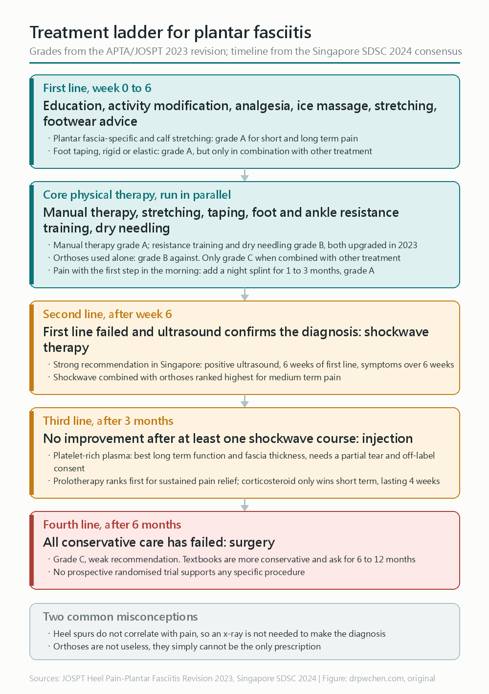
Treatment Ladder
- First-line (0–6 weeks): Mechanism education, activity modification, pain relief, ice massage, plantar fascia-specific stretch, footwear education; can add gastroc/soleus and Achilles stretching, antipronation taping (all Grade A)
SDSC 2024- Combined therapy (PT core): manual therapy (A) + stretching (A) + taping (A) + resistance training (B) + dry needling (B); insoles are only justified when used as an adjunct (C)
JOSPT CPG 2023- For first-step morning pain: Add night splint for 1–3 months (A)
JOSPT CPG 2023- No improvement at 6 weeks + positive ultrasound: ESWT (Grade A strong recommendation)
SDSC 2024- Still no improvement 3 months after at least one round of ESWT: Injection therapy — choose steroid for short-term pain relief, PRP/prolotherapy for long-term
- Symptoms lasting over 6 months with all conservative options failed: Surgery (Grade C, weak recommendation)
SDSC 2024Do not use insoles alone, and do not use therapeutic ultrasound to enhance stretching effects (both are recommendations against in the guidelines)
First-Line Care and Physical Therapy
- Ice, rest, NSAIDs, activity modification
- First-line combination explicitly listed in the guidelines (Grade A)
SDSC 2024- Physician explanation of biomechanical causes (education itself is a recommended item)
- Activity modification, pain relief, ice massage
- Plantar fascia-specific stretching
- Footwear education
- 2023 Guidelines grades for various interventions
JOSPT CPG 2023
| Intervention | Grade | Key Points |
|---|---|---|
| Manual therapy (joint + soft tissue) | A | Targets deficits in lower extremity joint ROM and flexibility |
| Stretching (PF-specific + gastroc/soleus) | A | Validated for short- and long-term pain relief and functional improvement |
| Taping (rigid or elastic) | A | Must be used with other treatments, for short-term pain relief |
| Night splint | A | For those with first-step morning pain, 1–3 month course |
| Therapeutic ultrasound | A (against) | Should not be used to enhance the effects of stretching |
| Resistance training (foot & ankle muscles) | B | Was F in the 2014 version, upgraded this time |
| Dry needling (calf and plantar MTrPs) | B | Was F in the 2014 version, reversed this time |
| Low-level laser | B | Short-term pain relief; 2–3 points, 904 nm at least 2 J/point |
| Insoles (used alone) | B (against) | Standalone use not recommended; was originally A in 2014 |
| Insoles (adjunct to other treatments) | C | Can be used to reduce pain and improve function |
| Phonophoresis + ketoprofen gel | C | Pain reduction |
| Electrotherapy | D | Prioritize manual therapy/stretching/insoles over electrotherapy; iontophoresis as second-line |
| Weight education and nutrition referral | E | Expert opinion |
- Evidence basis for the downgrade of insoles
Guimarães MA (in JOSPT CPG 2023)- Insole vs. control (sham or flat pad): 1–6 weeks P = .31, 7–12 weeks P = .06; neither showed significant differences
- Custom vs. prefabricated: 1–6 weeks P = .34, 7–12 weeks P = .72; also no difference
- However, when combined with ESWT and posterior chain stretching, the custom insole group had significantly better VAS scores at both 1 and 6 months (P = .0001)
- The conclusion is not that “insoles are useless”, but that insoles cannot serve as a standalone prescription
Exercise Prescription and Stretching
- Achilles tendon stretch
- Must first differentiate whether the Gastrocnemius or Soleus is tight (Silfverskiöld test)
- Plantar fascia-specific stretch: Using the windlass mechanism, manually pull the toes into dorsiflexion
- Strengthen short plantarflexors: Towel curls with the toes, note this is an opponens movement, otherwise it engages the long plantarflexors in the calf
- The guidelines have upgraded “resistance training for foot and ankle muscles” from not recommended to Grade B
JOSPT CPG 2023
- The guidelines have upgraded “resistance training for foot and ankle muscles” from not recommended to Grade B
Biomechanics and Orthotics
- Insoles (must be combined with other treatments)
- Early tender point is at the calcaneal attachment on the medial side → the insole should be hollowed out there to avoid compression
- Elevating the heel by 5–10 mm can immediately reduce pain through two mechanisms
Ombregt Cyriax 3e Ch.58- Shortens the distance between the metatarsus and calcaneus → directly relaxes the plantar fascia
- Removes Achilles tendon tension → indirectly relaxes the fascia
- The top surface of the heel must be flat; wedge shapes (women’s shoes) don’t work; women’s shoes need an extra anterior-thick wedge to level them
- Use an anterior rocker → the anterior MPJ doesn’t need dorsiflexion → reduces the windlass effect
- Kinesio taping: The guidelines have lumped both rigid and elastic taping together as Grade A (must be used as adjuncts)
JOSPT CPG 2023 - Night splint: Keeps the foot at 90 degrees at night
- Grade A, but the indication is restricted to those with “first-step morning pain”, for a course of 1–3 months
JOSPT CPG 2023 - ⚠️ For the 2023 version, no new studies were found for this item; the grade carried over from 2014
Trojian 2019 AFPtakes a reserved stance, considering that night splints and insoles have not been proven superior to placebo → both camps coexist, use only if the indication is correct- Practical limitation: Poor long-term compliance
- Grade A, but the indication is restricted to those with “first-step morning pain”, for a course of 1–3 months
- Double air-cushioned insole
- ⚠️ Low level of evidence:
Eun 2020 Biomed Res Intis a retrospective chart review (n = 40), not an RCT
- ⚠️ Low level of evidence:
- Flip-flops: May be considered (to train intrinsic muscles), but you have to choose ones with arch support
Extracorporeal Shockwave Therapy
- Guideline indications (Grade A strong recommendation): Ultrasound confirmation + failure of 6 weeks of first-line treatment + symptoms lasting over 6 weeks
SDSC 2024 - You need to use high-dose focused ESWT for better efficacy; it’s about 70% effective for pain control
- Network meta-analysis: ESWT combined with insoles had the highest SUCRA (80.3) for medium-term pain relief
Babatunde 2019 (in JOSPT CPG 2023)- Same analysis conclusion: No single treatment is superior to the others; CSI (alone or combined with exercise) and ESWT rank highest
- A level II RCT: At 24–48 weeks long-term follow-up, custom insoles were superior to ESWT in pain and function
Çaglar Okur (in JOSPT CPG 2023) - ⚠️ The JOSPT guidelines did not grade ESWT, on the grounds that it is considered outside the PT scope of practice, which does not mean the evidence is insufficient
Injection Therapies
Timing of Use
- PRP: Use only when “no improvement 3 months after at least one round of ESWT” + “evidence of partial tear” + “informed of off-label use” (Grade B strong recommendation)
SDSC 2024 - Perifascial corticosteroid injection: Ultrasound-guided, reserved for those with persistent severe pain who failed other conservative treatments (Grade B weak recommendation); pain relief only lasts up to 4 weeks
SDSC 2024 - Post-injection follow-up: Once at 2–4 weeks and once at 3–4 months; ultrasound can be used to track lesion changes
SDSC 2024 - Return-to-play criteria: At least 2 weeks post-injection, VAS below 2/10, lesion turned isoechoic or hyperechoic, and adapted to new insoles
SDSC 2024
PRP
- Level 1 evidence (Supported by consecutive MAs in 2024–26)
Zuo 2025 AJPMR MA24 RCTs n = 1,653: PRP vs. steroid, pain was better at 3 and 6 months (P = .03, P < .001), AOFAS better at 6/12 months- ⚠️ Pain at 1 month and 12 months showed “no significant difference” (P = .12 and P = .08), steroids were not superior
Tien 2026 Sci Rep NMA63 RCTs n = 4,170: PRP yielded the best improvement in long-term function and plantar fascia thicknessHohmann 2021 AJSM MA15 papers: No difference at 1 month, PRP had better pain scores at 3/6/12 months
- Indications: Failed conservative treatment for over 3 months, confirmed plantar fascia thickening
- US-guided injection into the hypoechoic lesion at the plantar fascia origin
Dextrose Prolotherapy
- Level 1-2 evidence (Complementary to PRP: pain relief vs. function)
Tien 2026 Sci Rep NMA: Prolotherapy ranks highest for sustained pain relief- ⚠️ The original text says prolotherapy, without specifically limiting it to dextrose
- The same analysis also notes that BoNT-A is best for short-term pain relief and thickness, while steroids are best for short-term function
Qafesha 2026 J Foot Ankle Res MA5 RCTs + 2 cohorts n = 567: CS is better at 1 month (VAS, FFI, thickness); DPT’s FFI is better at 3 months (P = .003)Asheghan 2021 Foot Ankle SurgRCT n = 59: rESWT × 3 vs. USG 20% dextrose 2 mL × 2, efficacy is comparable (only FAAM-sport leaned toward ESWT)Baykut 2025 Arch Orthop Trauma SurgRCT n = 70: fESWT vs. 15% dextrose 3 mL × 3, 12-week medium-term VAS leaned toward prolo
- Concentration: 15–20% dextrose, US-guided to the plantar fascia lesion
HVIGI
- ⚠️ Evidence is relatively less than for Achilles and Patellar; used on a case-by-case basis, no RCT-level evidence
Traditional Approach with Triamcinolone
- Excellent short-term pain relief but prone to relapse; see complications section above for risks
- Injection outcome depends entirely on accuracy; you must carefully locate the extent of the lesion before inserting the needle
Ombregt Cyriax 3e Ch.58 - Injection technique
Ombregt Cyriax 3e Ch.58- Patient prone, knee bent at 90 degrees, maintain dorsiflexion to tense the fascia (assistant holds it or you press it with the back of your injecting hand)
- Physician presses the tender point with their thumb, wrapping the other hand around the heel
- Needle: 2 mL triamcinolone, 4–5 cm long needle
- Enter from the medial edge of the fascia, about 3 cm away from the lesion (the skin over the tender point is too thick to disinfect well)
- Insert the needle at an oblique angle parallel to the fascia fibers, aiming straight for the tenoperiosteal junction
- Pass through the fascia and hit the bone → infiltrate at the tenoperiosteal border
- It will hurt a lot, and they might be unable to stand or walk for 24–48 hours due to the pain; you must inform them beforehand
- Follow-up at 14 days; if not fully relieved, give another injection
- Post-injection must be paired with heel elevation and short plantar flexor strengthening, or it will likely relapse
Ombregt Cyriax 3e Ch.58
Summary of Injection Choices
- Acute, severe pain (within 1 month): Steroid provides faster short-term relief, but the difference compared to PRP at 1 month is actually not significant
- Subacute to chronic (1–3 months): One steroid shot → re-evaluate
- Chronic, failed conservative treatment (over 3 months): PRP (strongest long-term evidence) or dextrose prolotherapy
- Concurrent plantar fascia thickening and structural pathology: PRP preferred
Surgical Options
- Timing: Symptoms over 6 months and conservative treatment failed (Grade C, weak recommendation)
SDSC 2024- Textbooks are more conservative: Consider only after at least 6 months, or even 12 months of failed non-surgical treatment
DeLee 5e Ch.120 - ⚠️ There are currently no prospective RCTs supporting any specific surgical procedure
DeLee 5e Ch.120
- Textbooks are more conservative: Consider only after at least 6 months, or even 12 months of failed non-surgical treatment
- Plantar fasciotomy (open or endoscopic)
- Tenotomy of the fascia origin at the heel under local anesthesia, resting for a few days post-op and strengthening the short plantarflexors
Ombregt Cyriax 3e Ch.58 - Open provides a full field of view and allows simultaneous decompression of the first branch of the lateral plantar nerve, but has higher wound complication rates and longer time to return to work
- Endoscopic has a faster recovery, but a smaller field of view, potential for under-resection, and inability to expose the lateral plantar artery and nerve, carrying a risk of injury
DeLee 5e Ch.120
- Tenotomy of the fascia origin at the heel under local anesthesia, resting for a few days post-op and strengthening the short plantarflexors
- Gastrocnemius recession
- Used for those with isolated gastrocnemius tightness with a positive Silfverskiöld test, to release plantar fascia tension
DeLee 5e Ch.120 - ⚠️ Still requires more studies to validate
- Used for those with isolated gastrocnemius tightness with a positive Silfverskiöld test, to release plantar fascia tension
- Spur excision: The spur has little relationship with symptoms, so resecting the spur is not the surgical goal
The Workflow Behind This Note
This note was originally my personal compilation of a common outpatient complaint. To write it up into a public version this time, I walked through the sources all over again.
Materials used
- Society guidelines and consensus: The American Physical Therapy Association’s JOSPT Heel Pain–Plantar Fasciitis Revision 2023 (the latest and most comprehensive one on this topic, including recommendation grades for various interventions), the 2024 18-statement consensus from Singapore’s SingHealth Duke-NUS Sports Medicine Centre, the 2018 American College of Foot and Ankle Surgeons consensus statement on infracalcaneal heel pain, and the older 2014 JOSPT guidelines for version comparison
- Textbooks: Cyriax Orthopaedic Medicine (Ombregt 3e), DeLee’s Orthopaedic Sports Medicine 5e, Giangarra’s Clinical Orthopaedic Rehabilitation 4e, Martinoli’s Musculoskeletal Ultrasound, Stecco’s Functional Atlas of the Human Fascial System, Bradley’s Atlas of Musculoskeletal Ultrasound Anatomy, Brukner & Khan’s Clinical Sports Medicine
- Primary literature: Stecco 2013’s cadaveric and MRI anatomical study, Trojian 2019’s family medicine review, four meta-analyses on injection therapies (Zuo 2025, Tien 2026, Hohmann 2021, Qafesha 2026) and two randomized trials (Asheghan, Baykut), as well as Tsai 1999 on heel fat pad mechanics
Tools used
audit_note.py— a note formatting audit script I wrote myself to check citation placements, images, and heading structures- PubMed MCP — to look up society guidelines and fill in volume/issue/page numbers, DOIs, and PMIDs for the bibliography, and read through abstracts one by one to verify numbers
- textbook_search — to do semantic search against my local textbook markdown indexes
- OpenEvidence — to do independent cross-searches on the controversies around insoles and night splints
- Python/PIL plotting scripts I wrote myself — for the treatment ladder diagram and the cover image (
scripts/figures/) - Hugo’s bilingual workflow
i18n_sync.py— machine translating the English version first, then manually proofreading the medical terms again
This workflow itself is written up in textbook-to-note. If you also have a Claude or Codex subscription, you can hand all this mechanical labor of verifying and compiling over to it, and leave the judgment to yourself. You can start with Getting Started with AI: Installation and the First Step and How to Talk to AI Agents.
About this version
The professional section is taken straight from my own clinical notes and wasn’t rewritten for the blog. The citation format follows my note-taking style (book title + chapter, or author + year, with the full bibliography below). Copyrighted images from textbooks and journals are not included here; the diagrams are redrawn by me.
If there are any misunderstandings, I welcome your corrections.
Reference
Guidelines and Consensus
- Koc TA Jr, Bise CG, Neville C, Carreira D, Martin RL, McDonough CM. Heel Pain – Plantar Fasciitis: Revision 2023. J Orthop Sports Phys Ther. 2023;53(12):CPG1-CPG39. doi:10.2519/jospt.2023.0303. PMID 38037331
- Tan VAK, Tan CC, Yeo NEM, et al. Consensus statements and guideline for the diagnosis and management of plantar fasciitis in Singapore. Ann Acad Med Singap. 2024;53(2):101-112. doi:10.47102/annals-acadmedsg.2023211. PMID 38920234
- Schneider HP, Baca JM, Carpenter BB, Dayton PD, Fleischer AE, Sachs BD. American College of Foot and Ankle Surgeons Clinical Consensus Statement: Diagnosis and Treatment of Adult Acquired Infracalcaneal Heel Pain. J Foot Ankle Surg. 2018;57(2):370-381. doi:10.1053/j.jfas.2017.10.018. PMID 29284574
- Martin RL, Davenport TE, Reischl SF, et al. Heel pain-plantar fasciitis: revision 2014. J Orthop Sports Phys Ther. 2014;44(11):A1-33. doi:10.2519/jospt.2014.0303. PMID 25361863
Journals
- Stecco C, Corradin M, Macchi V, et al. Plantar fascia anatomy and its relationship with Achilles tendon and paratenon. J Anat. 2013;223(6):665-676. doi:10.1111/joa.12111. PMID 24028383
- Trojian T, Tucker AK. Plantar Fasciitis. Am Fam Physician. 2019;99(12):744-750. PMID 31194492
- Zuo A, Gao C, Jia Q, et al. Platelet-Rich Plasma Versus Corticosteroids in the Treatment of Plantar Fasciitis: A Systematic Review and Meta-analysis. Am J Phys Med Rehabil. 2025;104(7):613-621. doi:10.1097/PHM.0000000000002677. PMID 39778212
- Tien CH, Chiu MC, Shen YL, Ko YC, Lee JJ. Comparative effectiveness of minimally invasive therapies for plantar fasciitis: a systematic review and network meta-analysis. Sci Rep. 2026;16(1). doi:10.1038/s41598-026-40038-z. PMID 41691098
- Hohmann E, Tetsworth K, Glatt V. Platelet-Rich Plasma Versus Corticosteroids for the Treatment of Plantar Fasciitis: A Systematic Review and Meta-analysis. Am J Sports Med. 2021;49(5):1381-1393. doi:10.1177/0363546520937293. PMID 32822236
- Qafesha RM, Ishreiteh HA, Nassourah AL, Tawil OI, Mashaly D. Efficacy and Safety of Dextrose Prolotherapy Versus Corticosteroid Injections in Plantar Fasciitis: A Systematic Review and Meta-Analysis. J Foot Ankle Res. 2026;19(1):e70135. doi:10.1002/jfa2.70135. PMID 41703400
- Asheghan M, Hashemi SE, Hollisaz MT, et al. Dextrose prolotherapy versus radial extracorporeal shock wave therapy in the treatment of chronic plantar fasciitis: A randomized, controlled clinical trial. Foot Ankle Surg. 2021;27(6):643-649. doi:10.1016/j.fas.2020.08.008. PMID 32919897
- Baykut AU, Bayraktar HEN, Arslan HB, Yalçın E, Poyraz İ. Chronic plantar fasciitis: which is more effective? Prolotherapy/extracorporeal shock wave therapy? A randomized controlled trial. Arch Orthop Trauma Surg. 2025;145(1):242. doi:10.1007/s00402-025-05859-z. PMID 40220030
- Eun SS, Chachan S, Lee SH. Effectiveness of a Double Air-Cushioned Shoe Compared with Physiotherapy in the Treatment of Plantar Fasciitis. Biomed Res Int. 2020;2020:9468302. doi:10.1155/2020/9468302. PMID 32337285
- Tsai WC, Chiu MF, Wang CL, Tang FT, Wong MK. The mechanical properties of the heel pad in unilateral plantar heel pain syndrome. Foot Ankle Int. 1999;20(10):663-668. doi:10.1177/107110079902001010. PMID 10541000
- Tedeschi R. Baxter’s nerve: the hidden culprit of chronic heel pain. Neurol Sci. 2025;46(9):4685-4689. doi:10.1007/s10072-025-08253-0. PMID 40418415
Textbooks
- Ombregt Cyriax 3e Ch.58 — Ombregt L. A System of Orthopaedic Medicine. 3rd ed. Elsevier; 2013. Disorders of the ankle and subtalar joints, p.770-771
- DeLee 5e Ch.120 — DeLee, Drez, & Miller’s Orthopaedic Sports Medicine. 5th ed. Elsevier; 2020. Heel Pain and Plantar Fasciitis
- Giangarra Clinical Ortho Rehab 4e Ch.43 — Giangarra CE, Manske RC. Clinical Orthopaedic Rehabilitation. 4th ed. Elsevier; 2018
- Martinoli MSK US 1e Ch.17 — Bianchi S, Martinoli C. Musculoskeletal Ultrasound. 1st ed. Springer; 2007. Foot
- Stecco Functional Atlas 1e — Stecco C. Functional Atlas of the Human Fascial System. 1st ed. Elsevier; 2015
- Bradley Atlas MSK US 2e — Bradley M, O’Donnell P. Atlas of Musculoskeletal Ultrasound Anatomy. 2nd ed. Cambridge University Press; 2009
- Brukner 5e Ch.43 — Brukner & Khan’s Clinical Sports Medicine. 5th ed. Foot pain