Bottom line up front: If your child has a painful bump just below the kneecap, it’s not growing pains. It has a name: Osgood-Schlatter, also known as tibial tubercle apophysitis.
And contrary to what many people have heard—it doesn’t “completely go away when you grow up." I’m writing this for parents who have 10- to 15-year-olds currently running and jumping in sports.
How to tell if it’s growing pains
Growing pains usually happen in both legs, ache in the evening or middle of the night, and have no clear point of tenderness. By the next morning, it’s like nothing happened.
Osgood-Schlatter is the exact opposite:
- The pain is pinpoint. It’s exactly on the prominent bump about two or three centimeters below the kneecap, and pressing it makes them jump.
- It hurts during exercise and improves with rest. Running, jumping, taking the stairs, and kneeling make it particularly obvious.
- That bone will get more prominent, feeling larger than the one on the other leg.
- It’s often just one leg, or one side is much worse than the other.
There’s a quick test you can do at home: have your child sit, block their shin with your hand, and ask them to kick their leg straight against your resistance. If this “forceful straightening” triggers pain at that spot, it’s highly likely.
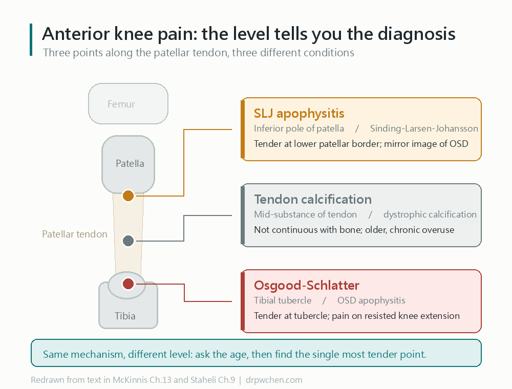
Another thing that often gets confused: with anterior knee pain, the height of the pain means a different disease. The same ligament has three different diagnoses corresponding to three locations from top to bottom.
Why does it happen?
When kids shoot up during puberty, their bones grow faster than their muscles. The muscles on the front of the thigh become relatively too tight, and all their pulling force concentrates on that “not-yet-hardened” growth plate below the kneecap.
With every jump and landing, that force pulls. Pull it too many times, and that growth plate gets inflamed, swollen, and stretched out—that’s where the bump comes from.
So it almost exclusively happens at a specific age: boys aged 12 to 15, girls aged 8 to 13 (girls are two years earlier because they develop earlier). Once the growth plate closes, new cases stop appearing.
About 1 in every 5 to 10 athletic kids gets it, which is much higher than in inactive kids.
Do we need an X-ray?
Usually, no. This is a condition you diagnose by “feel”: the right location, tenderness, and pain on forced extension are enough.
I order X-rays in these situations: if it’s purely one-sided and very atypical, if the pain wakes them up at night, if there’s fever or weight loss, if the swelling is more than you’d expect, or if I suspect something else that needs to be ruled out.
There’s one more situation where the threshold should be lower: if the child was referred in to a specialist by another doctor. The same knee pain carries a different distribution of underlying causes in a general clinic versus a referral center, so what needs ruling out deserves to be ruled out more actively.
Why does this matter? Bone tumors in children are rare, but they cluster around the knee and peak at age 14 — almost exactly the same location and age range as this condition. In published series, about a third of osteosarcomas were initially called tendonitis, with a median delay of over two months to diagnosis. And only about 20% of those children had night pain, so “it doesn’t hurt at night” is not reassurance.
This is also where ultrasound can’t help: ultrasound cannot see inside bone. It shows tendons, bursae, and cartilage well, but lesions in the marrow sit behind the cortical surface and simply aren’t visible. So “the ultrasound looked clean” is not an exclusion. Ruling out a tumor or infection still comes back to the X-ray.
One warning: seeing the bone look “fragmented” on an X-ray doesn’t necessarily mean there’s a disease. Some kids just naturally develop that way. So you can’t make the diagnosis just by looking at the film; it has to match the symptoms. Ultrasound has the same problem, and it’s harder to spot — in one follow-up study of young tennis players, 8 had ultrasound findings that looked like classic Osgood-Schlatter, and 7 of them had no pain at all.
The most common question: Should they stop sports completely?
No, and I don’t recommend it.
There’s actually a consensus in the medical community about this—surveys show nearly 80% of doctors are against complete rest for these kids. The right approach is “relative rest”:
- Keep moving within a tolerable range of pain. If it hurts, dial it back; if it doesn’t, slowly ramp it back up.
- First, avoid the most aggravating moves: squats, kneeling, and riding a bike with the seat too low.
- When it’s really painful, cut back on intense sports for a short term (about 4 to 8 weeks), but don’t just lie there doing nothing.
Complete rest has two downsides: muscles atrophy, making it even easier to hurt again when they return to sports; and the child loses their exercise habits and peer group, which costs them more than the knee pain itself.
Four things you can do at home
- Stretch the front of the thigh: This is the most useful of all treatments. Stand and pull the heel toward the glutes, keeping the knee pointing down rather than flaring out. Hold for 30 seconds. Stretch the back (hamstrings) while you’re at it.
- Infrapatellar strap: That thin band you strap just below the kneecap. Research has measured that when running, it actually reduces the force of the thigh muscle pulling on the tubercle. It’s not a placebo.
- Ice: 15 to 20 minutes after exercise. It helps with the pain, but you should know it won’t speed up healing.
- Knee pads: If your child plays volleyball, soccer, or sports involving kneeling and impact, add a pad to protect that bump.
Training volume red lines (with specific numbers)
I think this part is the most practical for parents. The sports medicine society’s consensus gives a few numbers you can check against right away:
Training volume thresholds to watch out for
- Weekly training hours exceeding the child’s age (e.g., a 13-year-old training over 13 hours a week)
- Over 16 hours of intensive training per week
- Training in the same single sport for more than 8 months a year (essentially year-round)
- Specializing in only one sport before age 12, crowding out other sports and free play
Hitting all three—“year-round + single sport + prepubescent”—is called “early sport specialization,” and it’s a recognized risk factor for overuse injuries.
Conversely, letting kids play multiple sports and keeping an off-season every year is prevention in itself. And evidence shows this won’t make them any less competitive.
One thing you shouldn’t do
Do not inject steroids there.
While the literature doesn’t list it as an “absolute contraindication,” textbook phrasing is “not recommended,” and for good reason: there is no research proving it works, yet it’s known to risk subcutaneous tissue atrophy and tendon rupture—and the injection site is right next to an active growth area.
Will it heal? The honest version
Traditional textbook teaching says “over 90% will resolve on their own as they grow up.” That’s half true.
What resolves is the pain and swelling. It usually eases off slowly over 12 to 24 months, right around when the growth plates close.
What won’t disappear is the bump. And if a small, independent bone fragment formed inside back then, it likely won’t be reabsorbed and will stay in the tendon forever.
There is a heavyweight study from Denmark that tracked down adults who had visited the hospital for this disease between 1977 and 2020. The results:
- 85% still had the bump
- 73% still had pain or trouble at that spot in adulthood
- Their sports performance and quality of life scores were lower than the general population
(To be fair: this group was severe enough to require referral back in the day, so the percentages skew high. The average kid won’t have it this bad.)
But this study also gave us a crucial message: the longer the symptoms dragged on and the worse the pain was back then, the worse their adult knees fared. So “just tough it out” is not a good strategy. Addressing training volume and flexibility early on actually matters.
When you absolutely must see a doctor
Don’t wait if these happen
- They can’t lift their leg straight up while lying down, or the bone suddenly gets very swollen or out of place—the bone might have pulled off, and it needs same-day treatment.
- They start walking with the foot turned out, or the hip doesn’t rotate freely—knee pain is sometimes referred from the hip, and that needs an X-ray of the pelvis, not the knee.
- The pain wakes them at night, they have fever or weight loss, or it’s strictly one-sided and atypical—we need to rule out infection or tumors.
- The swelling is clearly worse than you’d expect.
- They’ve done the treatments faithfully for two to three months with zero improvement.
As for adults whose bone fragments are still constantly hurting, there are surgical options (removing the fragment, shaving down the bump) with good results. But that’s a conversation we only have after 6 to 12 months of conservative treatment has failed.
Clinical Pearls
- OSD is a traction apophysitis of the tibial tubercle; location is the core of the differential diagnosis — the tubercle is OSD, the inferior pole of the patella is SLJ, and the mid-substance of the tendon is simple calcification.
- Diagnosis is clinical (tubercle tenderness + pain on resisted knee extension); X-rays are only used for unilateral/atypical presentations to rule out avulsion, tumors, or infection.
- But the population sets the threshold: typical presentations in general practice need no imaging; for referred patients or in a referral center, radiography takes priority over ultrasound — 31% of osteosarcomas are initially labeled tendonitis, and their site and age overlap OSD (
Widhe 2000 via Staheli), while ultrasound is blinded to intramedullary lesions by cortical acoustic shadowing (ElMiedany Peds Ch.4,Beggs Ch.9).- Ultrasound alone cannot diagnose OSD: the classic “OSD-like” appearance is common in asymptomatic knees (7 of 8 players pain-free,
Ducher 2010), and normal ossification itself is staged, with competing classification schemes.- “Self-limiting” needs a correction: soft tissue swelling and pain usually resolve in 12–24 months with growth plate closure, but once a loose ossicle forms, it mostly will not absorb and disappear. An adult cohort showed 73% still had residual discomfort (
Krommes 2025).- Conservative management success rate is >90%; steroid injection is not recommended (risk of subcutaneous atrophy and tendon rupture) — textbook phrasing is “not recommended,” not formally listed as a contraindication.
- Refractory ladder: dextrose prolotherapy (conflicting RCT evidence) → ESWT (weak evidence, traditionally a relative contraindication in children, use caution) → ossicle excision + tubercleplasty after skeletal maturity (good outcomes).
Abbreviations
| Abbreviation | Full Term |
|---|---|
| OSD | Osgood-Schlatter disease |
| SLJ | Sinding-Larsen-Johansson disease |
| ESWT | Extracorporeal shockwave therapy |
| EFD | Energy flux density |
| TT | Tibial tubercle / tuberosity |
Disease Background
Definition
- Traction apophysitis of the tibial tubercle secondary ossification center: an overuse injury caused by repetitive traction from the patellar tendon (
Staheli Ch.9,Cuccurullo Ch.122). - The lesion is at the distal insertion of the patellar tendon (tibial side), making it a symmetrical counterpart to SLJ at the proximal insertion (inferior pole of the patella) (
McKinnis Ch.13). - Symptomatic presentation persisting into adulthood is termed unresolved OSD (also known as a Mital lesion) (
Mital 1980).
Epidemiology
- Predominantly occurs during the pubertal growth spurt: boys aged 12–15, girls aged 8–13 (girls present 2 years earlier) (
Miranda-Comas Ch.16,Staheli Ch.9).- No new onset after growth plate closure; only appears before closure.
- Prevalence in athletic populations is 10–21%, significantly higher than in non-athletes (
DeLee Ch.137,Miranda-Comas Ch.16). - Bilateral in 20–50% of cases (mostly unilateral, though Cuccurullo reports up to 50% bilateral) (
Cuccurullo Ch.122). - Highest risk in sports involving running, jumping, kneeling, and kicking.
Pathophysiology
- Discrepant growth rates between bone and soft tissues: during the growth spurt, the rectus femoris relatively shortens, increasing tension in the extensor mechanism (
DeLee Ch.137,Herring Tachdjians Ch.40). - Repetitive concentric/eccentric quadriceps contraction → multiple submaximal micro-tears / micro-avulsion fractures of the secondary ossification center cartilage → inflammation, ossification, and tubercle enlargement (
Frontera Ch.31,Cuccurullo Ch.122). - Rectus femoris tightness is a known risk factor and the theoretical basis for stretching therapies (
Herring Tachdjians Ch.40). - The association with patella alta is controversial: Staheli/Herring consider it related, but studies cited by DeLee found no link between patella alta, OSD, and tubercle avulsion (
Staheli Ch.9vsDeLee Ch.137).
Natural History Model
- Flowers & Bhadreshwar model: normal apophysis development subjected to excessive traction → inflammation → majority heal; a minority continue with inflammation and persistent loose ossicles (
Staheli Ch.9).
Complications List
- Rare, including (
Sarwark Ch.33,Staheli Ch.9):- Tibial tubercle avulsion fracture: chronic OSD weakens the apophysis; forceful quadriceps contraction can cause acute avulsion.
- Genu recurvatum: premature closure of the anterior proximal tibial epiphysis → anterior tibial bowing, knee hyperextension (severe but rare).
- Patellar tendon rupture (extremely rare).
- Persistent symptomatic ossicle (the most common long-term sequela).
Long-term Prognosis
- Soft tissue swelling and pain usually resolve over 12–24 months around growth plate closure (
Staheli Ch.9,Sarwark Ch.33,Herring Tachdjians Ch.40). - Residual tubercle prominence is common: most adults can still palpate the bump, usually asymptomatic.
- Not completely benign: Danish national cohort (adults with a history of adolescent OSD) (
Krommes 2025):- 85% still had tubercle prominence; 73% still experienced pain or trouble from that site.
- All six KOOS subscales were lower than normative estimates for healthy populations, with sports and quality of life showing the largest gaps (Cohen’s d > 0.8).
- Risk of jumper’s knee in adulthood OR 70.4 (95% CI 32.9–155.0) — OSD is highly associated with subsequent patellar tendinopathy.
- The longer the symptoms lasted and the stronger the pain, the worse the adult knee health (dose-response).
- Contrasted against classic textbook claims that “>90% resolve with maturity” — the discrepancy stems from sampling (Krommes used a secondary care referral cohort, skewing severe); but clinically, patients should be honestly informed that symptoms can persist.
- Poor prognostic factors: imaging evidence of tubercle fragmentation is correlated with residual prominence and long-term symptoms (
Herring Tachdjians Ch.40).
Clinical Assessment
Diagnostic Criteria
- Clinical diagnosis: localized tibial tubercle swelling + tenderness + pain provoked by resisted knee extension, without joint effusion (
Staheli Ch.9,Frontera Ch.31). - Meeting the clinical picture + soft tissue swelling is sufficient for diagnosis; routine imaging is not needed. X-rays are only ordered for unilateral, atypical cases, or when needing to rule out other lesions (
Staheli Ch.9).
Which population “no routine imaging” applies to — referred patients warrant a lower threshold
- The original context of that principle is the typical adolescent in general practice; for referred patients or in a referral center, the pre-test probability differs and the threshold for radiography should be lower.
Brukner Ch.44states the exception explicitly: severe anterior knee pain with more swelling than expected → radiograph to exclude bony tumour; the knee is precisely where osteogenic sarcoma clusters in the 10–30 age group.- Why a missed diagnosis is costly (
Widhe 2000 via Staheli):
- 31% of osteosarcomas are initially diagnosed as tendonitis (Ewing sarcoma 21%).
- Median diagnostic delay 9 weeks (Ewing 19 weeks); activity pain 85%, night pain only 21% — the absence of night pain does not exclude.
- Predilection sites around the knee (distal femur / proximal tibia), peak age 14 — almost completely overlapping OSD in both site and age.
- Ultrasound is structurally unable to help here: cortical acoustic shadowing means US cannot assess intramedullary lesions; for bone tumors it shows only the extraosseous soft tissue component, cortical integrity, and periosteal reaction (
ElMiedany Peds Ch.4,Beggs Ch.9).
- In other words, a “clean” ultrasound is not an exclusion — what needs ruling out sits exactly where US cannot look.
History Taking
- Progressive tibial tubercle pain, exacerbated by activity (running, jumping, kneeling, stairs) and relieved by rest.
- Parents often notice an antalgic gait (
DeLee Ch.137). - Recent history of a growth spurt.
Physical Examination
- Tibial tubercle tenderness + swelling + prominence; bony irregularities can be palpated in chronic cases (
DeLee Ch.137). - Tenderness possible over the distal 1/2 of the patellar tendon.
- Pain on resisted knee extension; quadriceps and hamstring tightness are common.
- No joint effusion (effusion should raise suspicion for other intra-articular pathologies).
X-ray Interpretation
- Lateral views are the most useful (
Cuccurullo Ch.122,Braddom 7e Ch.37):- Acute phase: anterior soft tissue swelling over the tibial tubercle, blurring of the infrapatellar fat pad (Hoffa) borders.
- After 3–4 weeks: tibial tubercle fragmentation / loose ossicles, sclerosis, tubercle enlargement.
- Soft tissue swelling >4 mm (compared to contralateral side) supports the diagnosis (
Frontera Ch.31).
- Pitfall: Simple apophyseal fragmentation can be a normal variant. It must correlate clinically; diagnosis should not be made solely on X-rays (
Herring Tachdjians Ch.40).
How to differentiate bone fragmentation seen within the patellar tendon?
The keys are location + continuity + age + point of maximal tenderness (
McKinnis Ch.13, see previous image for location reference):
- OSD: fragment/ossicle at the tibial tubercle (distal patellar tendon insertion), often accompanied by tubercle enlargement and deformity; tenderness is at the tubercle.
- SLJ: fragment/calcification at the inferior pole of the patella (proximal patellar tendon insertion), tenderness at the lower patellar border; shares the same mechanism as OSD as a symmetrical counterpart.
- Simple patellar tendon calcification (dystrophic / post-chronic tendinopathy): calcific deposits are in the mid-substance of the tendon, unconnected to bone, usually seen in older patients with chronic overuse.
- Will it disappear? Soft tissue swelling and symptoms will ease with maturity, but once an independently ossified ossicle / intra-tendinous calcification forms, it mostly “will not” auto-absorb. It will remain in the patellar tendon; patients with persistent symptoms have unresolved OSD and may require surgical excision (
Krommes 2025,Mital 1980).
Ultrasound
- Useful for early detection and follow-up (
Cuccurullo Ch.122,ElMiedany 2015 Ch.9):- Swelling of soft tissues and unossified cartilage, thickened distal patellar tendon with increased echogenicity.
- Ossification center fragmentation/irregularity, decreased internal echogenicity.
- Deep infrapatellar bursitis.
- Doppler: hypervascularity at the affected patellar tendon insertion.
- One 3-type scheme has Type I as igloo-like deformity of the tibial tubercle ossification + deep infrapatellar bursitis + patellar tendon changes (
ElMiedany Peds Ch.10).- ⚠️ Citation correction, 2026-08-08: this note previously attributed the scheme to
ElMiedany Ch.5(2015 MSK US in Rheumatic Diseases) — that was wrong. Ch.5 of that book is Psoriatic Arthritis and the volume contains no such scheme; its OSD content sits in Ch.9 and describes imaging features only. The actual source is a different book, Pediatric MSK Ultrasonography 2e (2019), Ch.10. - ⚠️ That chapter only states “classified into three types or stages” with references [16,17] and names no originator, and never spells out types II/III → whether it is simply a restatement of the Czyrny scheme below cannot be determined.
- ⚠️ Citation correction, 2026-08-08: this note previously attributed the scheme to
Three prerequisites for reading the ultrasound — OSD cannot be diagnosed on ultrasound alone
The “fragmentation may be a normal variant” pitfall from the X-ray section applies equally to ultrasound, and is harder to detect, because US visualizes unossified cartilage and normal development itself looks like a lesion.
① Locate which ossification stage is normal for this patient, or there is no baseline
- Normal tibial tuberosity development follows the Ehrenborg & Engfeldt four phases (
Ehrenborg via ElMiedany Peds Ch.15):
- Cartilage phase → Apophyseal phase (secondary ossification center appears) → Epiphyseal phase (coalescence with the proximal tibial epiphysis) → Bone phase (physeal closure).
- The stage itself tracks OSD risk: a cross-sectional ultrasound study of 731 Japanese basketball players / 1462 knees found OR 9.48 from stage C→A and OR 2.22 from A→E; females mature 1–2 years earlier (59.2% already at stage E by age 10, versus 8.0% of males) (
Kaneuchi 2018).
- ⚠️ Level 3 cross-sectional study in a single population (Japanese basketball players) — do not extrapolate as a diagnostic threshold.
② “OSD-like” findings are common in asymptomatic knees — the hardest point
- A 12-month longitudinal follow-up of tennis players found the appearance classically associated with OSD (large anechoic region ± ossicles, apophyseal irregularity) in 8 players, 7 of whom were entirely pain-free; the authors state plainly that “the imaging appearance that is classically interpreted as OSD was common in asymptomatic knees” (
Ducher 2010).
- And 62% of stage 1 appearances progressed to a mature enthesis within a year — that is a developmental stage, not a disease.
- Same position in
DeLee Ch.132: tubercle fragmentation is not in itself diagnostic and is seen in asymptomatic patients; that chapter also notes that for OSD, “ultrasound is not commonly performed”.③ Several classification schemes coexist, and none is validated
- Their axes are mutually incompatible and do not map onto each other:
Scheme Axis Source “igloo” 3 types Lesion morphology (Type I = igloo-like deformity + bursitis + tendon changes) ElMiedany Peds Ch.10(originator unnamed)Czyrny 3 types Delamination (1 of the internal ossification center / 2 tear or fracture of the epiphyseal portion / 3 delamination producing irregular tuberosity deformity) Czyrny via ElMiedany Peds Ch.19Ducher 3 stages Developmental stage (cartilage attachment / insertional cartilage / mature attachment) Ducher 2010(primary paper)Ehrenborg 4 phases Normal ossification, not a disease classification Ehrenborg via ElMiedany Peds Ch.15
- Chapters 10 and 19 of the same Pediatric MSK US 2e describe the “three types” in terms that do not line up (igloo-like deformity vs delamination); Ch.10 names no source, so a restatement of one scheme cannot be ruled out — the book simply doesn’t say.
- ⚠️ Always state which scheme and which chapter;
ElMiedanycovers two different books (2015 Rheumatic Diseases / 2019 Pediatric), and the bare surname points at the wrong one.Practical conclusion: in OSD, ultrasound belongs to follow-up and soft tissue differentiation (tendon, bursa, Doppler activity), not standalone diagnosis; diagnosis remains clinical, and exclusion questions go back to radiography.
MRI
- The most sensitive and specific modality (
Cuccurullo Ch.122,Martinoli Ch.19):- Tibial tubercle bone marrow edema, distal patellar tendon thickening, preinsertional tendinosis.
- Avulsion / bony fragmentation of the ossification center.
- Blurring of the inferior margin of the infrapatellar fat pad, distended deep infrapatellar bursa (common).
Staging and Classification
- Clinically, descriptive imaging staging is used more often than formal grading; see above for the competing 3-type ultrasound schemes.
- The key is distinguishing between active (immature, healable) vs. unresolved (persistent symptomatic ossicle post-maturity); the latter dictates surgical intervention (
Mital 1980).
Differential Diagnosis Table
| Differential | Lesion Location | Imaging Features | Key Distinguishing Factors |
|---|---|---|---|
| OSD | Tibial tubercle | Tubercle fragmentation/ossicle, enlargement | Tenderness at the tubercle; pain on resisted knee extension |
| SLJ | Inferior pole of the patella | Inferior pole fragmentation/calcification | Tenderness at the lower patellar border; symmetrical pathology with the same mechanism |
| Jumper’s knee (patellar tendinopathy) | Patellar tendon at the inferior pole (skeletally mature) | Tendon thickening, tendinosis, late-stage calcification | Skeletally mature athletes; not an apophyseal issue |
| Tibial tubercle avulsion fracture | Tibial tubercle | Acute displacement of bone fragment | SLR lags compared to contralateral side, history of acute trauma |
| Infrapatellar bursitis | Bursa deep to the patellar tendon | Bursal effusion | Localized swelling, no ossification center lesions |
| Pes anserine bursitis | Proximal medial tibia | Mostly normal | Tenderness is medial, not at the tubercle |
| SCFE (see below) | Hip (pain referred to knee) | Knee films normal; needs AP pelvis + frog-leg | Restricted hip internal rotation, obligate external rotation on flexion; age band overlaps OSD |
| Perthes | Hip (pain referred to knee) | Femoral head epiphysis collapse / fragmentation (may be normal early) | Ages 4–10, Trendelenburg / antalgic gait |
| Infection / Tumor | Variable | Osteolysis / periosteal reaction | Night pain, systemic symptoms, atypical presentation → rule out with imaging |
An adolescent with knee pain must have the hip assessed — the costliest miss in the OSD differential
- SCFE can present as knee pain alone, and knee pain may be the only symptom (
Sarwark Ch.20).
- Mechanism: pain referred along the anterior branch of the obturator nerve, which innervates both hip and knee (
Abdelgawad Ch.7).- The textbook wording is unusually firm: “It is important and medically indicated to evaluate the hip of the adolescent patient who presents with knee pain” (
Sarwark Ch.20); “Any child complaining of knee pain should have both the knee and hip joints assessed” (Abdelgawad Ch.7).- The age band overlaps OSD entirely: SCFE peaks at 11–12 in girls, 13–14 in boys (
Campbell Ch.14;Sarwark Ch.10lists 9–15).- Knee radiographs cannot show SCFE: you need AP pelvis plus frog-leg lateral, the latter being most sensitive to posterior slip (
Sarwark Ch.20,Staheli, SCFE).
- Always view the contralateral side: contralateral SCFE occurs in up to 25%.
- Signs: widened physis, reduced epiphyseal height, Klein line failing to intersect the epiphysis, metaphyseal rarefaction.
- Screening exam (either finding → order pelvis films): obligate external rotation on hip flexion (knee-to-shoulder sign), and reduced hip internal rotation.
- Perthes also presents as knee pain (ages 4–10) and may have normal early radiographs; high clinical suspicion warrants MRI / bone scan (
Sarwark Ch.10,Braddom 6e Ch.36).
Red Flags and Referral Timing
- Suspected avulsion fracture: inability to perform a straight-leg raise (SLR), visible widening/displacement of the tubercle → refer to pediatric orthopedics (urgent) (
Sarwark Ch.33). - Restricted hip internal rotation / obligate external rotation on hip flexion / out-toeing gait → AP pelvis + frog-leg lateral to rule out SCFE (not knee films); see the differential section (
Sarwark Ch.20). - Atypical / unilateral / night pain / systemic symptoms → image to rule out tumors or infection.
- Swelling clearly out of proportion to expectation → radiograph to exclude bony tumour (
Brukner Ch.44). - No improvement after weeks to months of conservative management → refer to pediatric sports medicine / orthopedics (
Sarwark Ch.33).
Other pediatric limb-pain red flags that mandate a radiograph
- Osteoid osteoma: ages 10–20, typically tibia or spine, night pain; radiographs may show a lucent nidus surrounded by sclerosis; bone scan is diagnostic (
Sarwark Ch.10).- Ewing sarcoma: ages 1–20, diaphyseal lesion with periosteal reaction; may cause fever and raised ESR → clinically and radiographically mimics osteomyelitis.
- One series found 1 Ewing sarcoma among 30 children with presumed osteomyelitis; taking a trocar bone biopsy at the same time as aspiration for culture is recommended (
Herring Tachdjians Ch.23).- Acute leukemia (ALL): bone and joint symptoms in 21–59% of children; in a review of 296 cases, 22% had bone pain and 18% had bone pain overshadowing other manifestations (
Herring Tachdjians Ch.23).
- The key trap: children with prominent bone pain frequently have near-normal hematologic values, which delays diagnosis; when suspected, request manual review of the peripheral smear for blasts.
- Radiographs may show metaphyseal lucent bands, osteopenia, lytic lesions, periosteal new bone; any child with vague musculoskeletal pain plus constitutional symptoms — with or without radiographic findings — warrants oncology consultation (
Abdelgawad Ch.15).- Osteomyelitis / septic arthritis: the chief value of plain films is to exclude tumor or fracture; obtain at least two planes (
Herring Tachdjians Ch.23).- Night pain cuts both ways: only 21% of osteosarcomas have night pain (activity pain 85%), so its absence does not exclude tumor; yet osteoid osteoma is almost always night pain. Night pain alone settles nothing — it is a trigger to image.
Clinical Management
Management Decision-Making
OSD Management Algorithm (Axes: Skeletal Maturity × Symptom Severity/Recalcitrance)
There are no formal society CPGs for OSD; the grades below are literature evidence levels, not society SoRs.
- Skeletally immature + typical presentation (vast majority) → patient education + relative rest + stretching, maintaining activity within tolerable limits → >90% success (
Herring Tachdjians Ch.40).
- Add infrapatellar strap, ice, and knee pads for symptom control.
- Recalcitrant to ≥3 months of conservative care with high athletic demands → consider dextrose prolotherapy (conflicting RCT evidence); or ESWT (weak evidence, traditionally a relative contraindication in children, use caution).
- Skeletally mature + symptomatic ossicle (unresolved OSD), failing 6–12 months of conservative care → ossicle excision + tubercleplasty (good outcomes).
- Suspicion of avulsion at any time (SLR lag vs. contralateral / tubercle displacement) → refer to orthopedics.
- Not recommended: steroid injection into the tibial tubercle or patellar tendon (risk of subcutaneous atrophy and tendon rupture) (
Staheli Ch.9,Herring Tachdjians Ch.40).
- Original texts use “not recommended / no longer recommended”; the literature does not formally list it as a contraindication. Opposition stems from clinical consensus + theoretical risk + lack of efficacy evidence; steroid injections fell into the “disagree” category in the Lyng 2020 survey (
Lyng 2020).- Clinical decision remains the same (do not inject), but in education and documentation, it shouldn’t be framed as an “absolute contraindication”.
Treatment Goals
- Control pain, maintain tolerable sports participation, protect the apophysis from avulsion, and await natural maturation.
Prevention and Training Volume Control
- No OSD-specific prevention guidelines exist; the following is society consensus for adolescent overuse injuries, which encompasses OSD (
AOSSM 2016). - Early sport specialization is defined by three conditions being met simultaneously (
AOSSM 2016):- Intensive training or competition for more than 8 months a year (almost year-round).
- Focusing on a single sport to the exclusion of other sports and free play.
- Occurring before puberty (around seventh grade / age 12).
- Two quantitative thresholds requiring close monitoring for overuse injury, burnout, and overtraining (
AOSSM 2016):- Weekly training hours exceeding the child’s age (e.g., >13 hours/week for a 13-year-old).
- Intensive training exceeding 16 hours per week.
- All adolescents (including non-athletes) can benefit from periodized integrative neuromuscular training (INT); specialized athletes especially need a dedicated INT phase (
AOSSM 2016). - Each sport has its own loading profile and corresponding overuse injuries — OSD correlates with running, jumping, kicking, and kneeling (
AOSSM 2016,DeLee Ch.137). - Concrete training volume example (safe training recommendations for tennis players aged 12 and older) (
AOSSM 2016):- Organized training under 12 hours per week, competition under 12 events per year.
- Injury prevention training at least 2 hours per week.
- Incorporate a second sport that has an off-season.
- ⚠️ The above are consensus statements (expert opinion level), not RCT-based evidence; and they are not validated against OSD endpoints specifically.
Conservative Treatment
Activity Modification / Relative Rest
- Relative rest (activity within tolerable discomfort), not strict bed rest — 78.9% of physicians disagree with complete rest.
- Evidence: Moderate — single-arm trial of education + activity modification + progressive strengthening improved self-reported outcomes (
Hansen 2023,Lyng 2020).
- Evidence: Moderate — single-arm trial of education + activity modification + progressive strengthening improved self-reported outcomes (
- Avoid deep knee flexion movements like squats, kneeling, or cycling with low seat height; for severe cases, restrict intense sports short-term (4–8 weeks) (
Cuccurullo Ch.122).
Acute Symptom Control
- Ice (15–20 mins, 2–3 times daily, especially post-exercise), short-term NSAIDs for pain relief (
Sarwark Ch.33,Herring Tachdjians Ch.40).- Evidence: Low — provides analgesia but does not accelerate healing or alter the disease course (
Miranda-Comas Ch.21).
- Evidence: Low — provides analgesia but does not accelerate healing or alter the disease course (
- Knee pads to protect the tubercle in impact-prone sports (e.g., volleyball, soccer).
Physical Therapy
- Quadriceps and hamstring stretching (targeting rectus femoris tightness) is the most useful treatment (
Staheli Ch.9,Herring Tachdjians Ch.40).- Evidence: Low to Moderate — largely based on narrative reviews and biomechanical rationale; case report showed return to competition after 2 months of kinesiotherapy + static stretching (
Gaweł 2021).
- Evidence: Low to Moderate — largely based on narrative reviews and biomechanical rationale; case report showed return to competition after 2 months of kinesiotherapy + static stretching (
- Addition of progressive quadriceps/gluteal strengthening, correcting landing mechanics, and addressing biomechanical factors like lumbo-pelvic control and subtalar pronation (
Miranda-Comas Ch.21).
Bracing
- Infrapatellar strap (placed over the patellar tendon between the patella and the tubercle) for offloading and analgesia (
Sarwark Ch.33,Herring Tachdjians Ch.40).- Evidence: Low — 2024 biomechanics study showed significant reductions in peak and cumulative quadriceps force during running (p < 0.001), primarily decreasing vastus forces; limited impact on rectus femoris forces, so patients with rectus tightness may benefit less (
Zhang 2024).
- Evidence: Low — 2024 biomechanics study showed significant reductions in peak and cumulative quadriceps force during running (p < 0.001), primarily decreasing vastus forces; limited impact on rectus femoris forces, so patients with rectus tightness may benefit less (
- Knee immobilizer occasionally used for 7–10 days in severe pain to reduce inflammation, removed daily for ROM exercises (
Staheli Ch.9,Abdelgawad Ch.8).
Emerging Therapies
Dextrose Prolotherapy
- 12.5–20% hyperosmolar dextrose injected into the tubercle / patellar tendon insertion, used for recalcitrant OSD.
- Conflicting RCT evidence, though all three trials reported good safety profiles without adverse events:
- Evidence: Conflicting — Topol 2011 (n=54) found dextrose superior to lidocaine and usual care, with a higher proportion of asymptomatic sports participation at 1 year (
Topol 2011); Wu 2022 (n=70, ultrasound-guided 12.5%) showed better VISA-P scores at 3, 6, and 12 months versus saline (Wu 2022). - Evidence: Negative — Nakase 2020 (43 knees, 20% dextrose) found no significant difference compared to saline; both groups improved over time (
Nakase 2020).
- Evidence: Conflicting — Topol 2011 (n=54) found dextrose superior to lidocaine and usual care, with a higher proportion of asymptomatic sports participation at 1 year (
- Discrepancies may stem from concentration differences (12.5% vs. 20%), use of ultrasound guidance, or sampling variations; not yet sufficient to become standard of care.
Extracorporeal Shockwave Therapy (ESWT)
- Mechanisms: disintegration of calcifications, hyperstimulation analgesia, neovascularization, alteration of cell permeability (
Bellew Ch.16). - Evidence: OSD-specific evidence is limited to case series; no RCTs or systematic reviews exist.
- Evidence: Very Low — multimodal analgesic adjunct in OSD (
Schmitz 2015,Lyng 2020).- Early high-energy focused ESWT was traditionally considered a relative contraindication in children due to concerns over growth plate damage in animal studies.
- Recent animal and case data suggest radial ESWT (rESWT) is relatively safe in immature bone.
- Evidence: Very Low — case series of 15 OSD patients receiving focused, low-energy ESWT reported no adverse events (
Shafshak 2023).
- Evidence: Very Low — multimodal analgesic adjunct in OSD (
- Suggested protocol parameters (no OSD-specific consensus; adopting general soft-tissue tendinopathy guidelines) (
Braddom 7e Ch.18):- 2000–3000 shocks per session, 3 consecutive sessions, once a week.
- EFD set at the upper limit of moderate intensity tolerated by the patient (low <0.12, high >0.12 mJ/mm² are common cutoffs; alternative definitions use low 0.08–0.28, medium 0.28–0.60, high >0.60 mJ/mm²).
- Ultrasound localization of lesion depth as the target.
- Contraindications: bleeding disorders, pacemakers, anticoagulation, pregnancy, acute injuries — along with “children” as a traditional relative contraindication. Therefore, its use in OSD should be restricted to recalcitrant cases or those nearing skeletal maturity, with fully informed consent (
Bellew Ch.16).
- Out-of-pocket reference (Taiwan): Not covered by National Health Insurance.
- Nationally announced range is approximately NT$1000–2000 per session.
Surgical Interventions
Ossicle Excision + Tubercleplasty
- Indications: skeletally mature + symptomatic ossicle (unresolved OSD), failing 6–12 months of conservative care, with persistent kneeling pain / activity limitations (
Sarwark Ch.33,Mital 1980). - Procedure: excision of loose ossicles and bursal tissue, often combined with distal patellar tendon debridement and tubercleplasty; can be performed open, arthroscopically, bursoscopically, or ultrasound-guided.
- Evidence: Moderate (consistently good in case series) — arthroscopic ossicle excision + tubercleplasty improved Kujala scores from 69.9 to 98.5, Lysholm from 76.3 to 99.5, with 100% returning to pre-injury sports (
Dimnjaković 2026); bursoscopic excision in military personnel improved Lysholm from 71 to 99, VAS from 6.5 to 0.9, with 17/18 satisfied (Eun 2015). - Evidence: Moderate — long-term follow-up of military personnel post-surgery showed 87% without daily limitations and 75% returning to pre-surgical activity levels (
Pihlajamaki via DeLee Ch.137). - ⚠️ Case series have small sample sizes (the former n=12, mean age 21.7 years, preoperative symptoms lasting an average of 77 months) and lack randomized controls.
- Evidence: Moderate (consistently good in case series) — arthroscopic ossicle excision + tubercleplasty improved Kujala scores from 69.9 to 98.5, Lysholm from 76.3 to 99.5, with 100% returning to pre-injury sports (
- Surgical complications cannot be ignored: out of 6 surgical studies reviewed, 5 reported complications ranging from 0–75.8%, including infection, hypertrophic scarring, and residual pain (
Ndjonko 2026).- Compared to 0–66.7% in non-surgical groups (mostly quadriceps atrophy and skin irritation) — the advantage of surgery lies in efficacy, not safety.
- A literature review (15 studies / 712 patients) covered non-surgical, injection, and surgical categories, with surgery reserved for recalcitrant mature cases (
Ndjonko 2026).- ⚠️ The article title claims to be a systematic review, but the authors self-described the design as a scoping review, Level of evidence 4; it should not be cited as high-level evidence.
Post-operative Rehabilitation
- Progressive quadriceps/hamstring strengthening and stretching, ROM restoration; return to play dictated by symptoms and muscle strength. Principles echo conservative rehabilitation, avoiding premature high-load traction on the tubercle.
Evidence Summary Table
| Intervention | Evidence | Conclusion | Key Studies |
|---|---|---|---|
| Education + activity modification + progressive strengthening (first-line) | Moderate | >90% success, treatment of choice | Hansen 2023, Lyng 2020 |
| Quadriceps/hamstring stretching | Low–Mod | Most useful conservative option; evidence mostly narrative | Gaweł 2021, Herring Tachdjians Ch.40 |
| Infrapatellar strap | Low | Reduces quadriceps load, provides analgesia; less benefit for rectus tightness | Zhang 2024 |
| NSAIDs / Ice | Low | Analgesic, does not alter disease course | Miranda-Comas Ch.21 |
| Dextrose prolotherapy | Conflicting | 2 positive RCTs, 1 negative RCT; safe | Topol 2011, Wu 2022, Nakase 2020 |
| ESWT (OSD) | Very Low | Case series only, no RCTs; rESWT is safer | Shafshak 2023, Schmitz 2015 |
| Steroid injection | No evidence of efficacy | Not recommended (subcutaneous atrophy, tendon rupture); not a formal contraindication | Staheli Ch.9, Herring Tachdjians Ch.40, Lyng 2020 |
| Ossicle excision + tubercleplasty | Moderate (case series) | Good outcomes and high return-to-play in unresolved OSD, but complications range 0–75.8% | Dimnjaković 2026, Eun 2015, Ndjonko 2026 |
| Training volume control / avoiding early sport specialization | Consensus (expert opinion) | Quantitative thresholds available for education; not validated against OSD endpoints | AOSSM 2016 |
Production Workflow for These Notes
Materials used
- Pediatric orthopedics and sports medicine textbooks: Tachdjian’s 6e, Staheli 4e, Sarwark, DeLee 5e, Braddom 7e, Cuccurullo 4e, Miranda-Comas, Frontera, Abdelgawad; imaging and ultrasound from McKinnis 5e, Martinoli, and ElMiedany
- Consensus statement: the AOSSM 2016 early sport specialization consensus (the source for the prevention and training-volume section)
- Primary literature: the Danish national cohort study on residual symptoms in adulthood (Krommes 2025), three dextrose prolotherapy RCTs (Topol 2011 / Nakase 2020 / Wu 2022), the surgical series (Eun 2015, Dimnjaković 2026, Mital 1980), and reviews of ESWT efficacy and safety
- Evidence on patient education design: Hansen 2023 on developing a leaflet for children, and Lyng 2020 on current clinical practice
Tools used
audit_note.py— my own note-format audit script- PubMed MCP — filling in volume/issue/page and PMIDs for the three 2025–2026 papers, and checking each paper’s stated study design and level of evidence
- textbook_search — semantic search over a local markdown index of my textbooks
- OpenEvidence — an independent search to sanity-check the treatment-recommendation wording
- Custom Python/PIL plotting scripts — the anterior knee pain location figure and the cover image, one of each per language (
scripts/figures/) - Hugo’s bilingual pipeline
i18n_sync.py— the English version is machine-translated first, then medical terminology is proofread by hand
This workflow itself became textbook-to-note. If you have a Claude or Codex subscription, this kind of mechanical verification and organizing work can be handed off, leaving the judgment to you. Getting started and talking to agents are good entry points.
About this version
The pro version is taken straight from my own clinical notes, unedited for the blog, keeping my original citation style (book + chapter, or author + year; full bibliography below). Copyrighted images from textbooks and journals aren’t included here — every figure is one I redrew myself.
Revised 2026-08-08: a physician raised two points, both valid, now incorporated into the text — (1) for pediatric limb pain in a referral population, radiography takes priority over ultrasound (new callout on which population “no routine imaging” applies to); (2) normal tibial tuberosity ossification is itself staged, and OSD ultrasound classification schemes differ between authors (new callout on the three prerequisites for reading the ultrasound). With thanks for the correction.
If I’ve gotten something wrong, corrections are welcome.
Reference
Books
- Staheli Ch.9 — Fundamentals of Pediatric Orthopedics, 4th ed, 2008
- Sarwark Ch.33 — Pediatric Orthopaedics and Sports Injuries: A Quick Reference Guide, 3rd ed, 2021
- Cuccurullo Ch.122 — Physical Medicine and Rehabilitation Board Review, 4th ed, 2020
- Herring Ch.40 — Tachdjian’s Pediatric Orthopaedics, 6th ed, 2022
- DeLee Ch.137 — DeLee & Drez’s Orthopaedic Sports Medicine, 5th ed, 2020
- Braddom Ch.18 / Ch.37 — Physical Medicine and Rehabilitation, 7th ed, 2021 (ESWT physical parameters, Lower Limb Pain)
- McKinnis Ch.13 — Fundamentals of Musculoskeletal Imaging, 5th ed, 2022 (OSD vs SLJ differential)
- Martinoli Ch.19 — Musculoskeletal Ultrasound, 1st ed, 2007
- Frontera Ch.31 — Clinical Sports Medicine, 1st ed, 2007
- Miranda-Comas Ch.16 / Ch.21 — Essential Sports Medicine, 2nd ed, 2021
- Bellew Ch.16 — Michlovitz’s Modalities for Therapeutic Intervention, 6th ed, 2016
- Abdelgawad Ch.8 — Pediatric Orthopedics and Sports Medicine, 2nd ed, 2020
- ElMiedany 2015 Ch.9 — Musculoskeletal Ultrasonography in Rheumatic Diseases, 1st ed, 2015 (Pediatric MSK Disorders; OSD imaging features, no classification scheme)
- ElMiedany Peds Ch.4 / Ch.10 / Ch.15 / Ch.19 — Pediatric Musculoskeletal Ultrasonography, 2nd ed, 2019 (Ch.4 limits of US for marrow, Ch.10 igloo three types, Ch.15 Ehrenborg four phases, Ch.19 Czyrny three types)
- ⚠️ Two different books, same editor; before 2026-08-08 this note misattributed the igloo scheme to “ElMiedany Ch.5” (that chapter is Psoriatic Arthritis)
- Beggs Ch.9 — Musculoskeletal Ultrasound, 1st ed, 2014 (limits of US in evaluating bone tumors)
- Brukner Ch.44 — Clinical Sports Medicine Vol.1, 5th ed, 2017 (The Younger Athlete)
- DeLee Ch.132 — DeLee & Drez’s Orthopaedic Sports Medicine, 5th ed, 2020 (Imaging Considerations in Skeletally Immature Athletes)
- Staheli, Malignant Bone Tumors / SCFE — Fundamentals of Pediatric Orthopedics, 4th ed, 2008 (initial misdiagnosis rates in pediatric bone tumors; knee pain warrants hip evaluation)
- Sarwark Ch.10 / Ch.20 — Pediatric Orthopaedics and Sports Injuries, 3rd ed, 2021 (The Limping Child differential table; SCFE)
- Abdelgawad Ch.7 / Ch.15 — Pediatric Orthopedics and Sports Medicine, 2nd ed, 2020 (SCFE referred-pain mechanism; leukemia imaging and referral)
- Herring Tachdjians Ch.23 — Tachdjian’s Pediatric Orthopaedics, 6th ed, 2022 (MSK infection; the ALL and Ewing misdiagnosis traps)
- Campbell Ch.14 — Physical Therapy for Children, 5th ed, 2017 (SCFE epidemiology and age of onset)
Papers
- LaPrade RF, Agel J, Baker J, et al. AOSSM Early Sport Specialization Consensus Statement. Orthop J Sports Med. 2016;4(4):2325967116644241. doi:10.1177/2325967116644241
- Krommes K, Bjerre A, Thorborg K, et al. Long-Term Knee Health in Adults with a History of Adolescent Osgood-Schlatter: A National Cohort Study of Patients in Secondary Care in Denmark 1977-2020. Sports Med. 2025;55(7):1769-1781. doi:10.1007/s40279-025-02214-5. PMID: 40439870
- Ndjonko LCM, Klein JH, Chakraborty Y, et al. Treatments for Osgood Schlatter Disease: A Systematic Review of the Literature. Orthop J Sports Med. 2026;14(3):23259671251387354. doi:10.1177/23259671251387354. PMID: 41788553
- Dimnjaković D, Plečko M, Đukić E, Bojanić I. Standard knee arthroscopy with free ossicle removal and tubercleplasty provides optimal results for unresolved Osgood-Schlatter disease. Knee. 2026;62:104498. doi:10.1016/j.knee.2026.104498. PMID: 42190337
- Topol GA, Podesta LA, Reeves KD, et al. Hyperosmolar Dextrose Injection for Recalcitrant Osgood-Schlatter Disease. Pediatrics. 2011;128(5):e1121-8. PMID: 21969284
- Nakase J, Oshima T, Takata Y, et al. No Superiority of Dextrose Injections Over Placebo Injections for Osgood-Schlatter Disease. Arch Orthop Trauma Surg. 2020;140(2). PMID: 31713082
- Wu Z, Tu X, Tu Z. Hyperosmolar Dextrose Injection for Osgood-Schlatter Disease: A Double-Blind, Randomized Controlled Trial. Arch Orthop Trauma Surg. 2022;142(9). PMID: 34673998
- Lyng KD, Rathleff MS, Dean BJF, et al. Current management strategies in Osgood Schlatter: A cross-sectional mixed-method study. Scand J Med Sci Sports. 2020;30(10):1985-1991. doi:10.1111/sms.13751. PMID: 32562293
- Hansen R, Rathleff MS, Lundgaard-Nielsen M, Holden S. Development of an informative leaflet for children with Osgood-Schlatter disease. Scand J Med Sci Sports. 2023;33(12). PMID: 37726948
- Gaweł E, Zwierzchowska A. Therapeutic Interventions in Osgood-Schlatter Disease: A Case Report. Medicine (Baltimore). 2021;100(50):e28257. PMID: 34918694
- Zhang X, Ren W, Wang X, et al. Quantitative Analysis of Quadriceps Forces in Adolescent Females During Running With Infrapatellar Straps. J Sports Sci Med. 2024;23(4):787. PMID: 39649570
- Shafshak TS, Amer MA. Extracorporeal shockwave therapy in Osgood-Schlatter disease (case series). J Orthop Surg Res. 2023. PMID: 37608382
- Schmitz C, Császár NB, Milz S, et al. Efficacy and Safety of Extracorporeal Shock Wave Therapy for Orthopedic Conditions. Br Med Bull. 2015;116(1):115-38. PMID: 26585999
- Eun SS, Lee SA, Kumar R, et al. Direct Bursoscopic Ossicle Resection in Young and Active Patients With Unresolved Osgood-Schlatter Disease. Arthroscopy. 2015;31(3). PMID: 25442658
- Mital MA, Matza RA, Cohen J. The So-Called Unresolved Osgood-Schlatter Lesion. J Bone Joint Surg Am. 1980;62(5). PMID: 7391096
- Kaneuchi Y, Otoshi K, Hakozaki M, et al. Bony Maturity of the Tibial Tuberosity With Regard to Age and Sex and Its Relationship to Pathogenesis of Osgood-Schlatter Disease: An Ultrasonographic Study. Orthop J Sports Med. 2018;6(1):2325967117749184. doi:10.1177/2325967117749184. PMID: 29344541
- Ducher G, Cook J, Spurrier D, et al. Ultrasound Imaging of the Patellar Tendon Attachment to the Tibia During Puberty: A 12-Month Follow-Up in Tennis Players. Scand J Med Sci Sports. 2010;20(1):e35-40. doi:10.1111/j.1600-0838.2009.00889.x. PMID: 19486483
- Widhe B, Widhe T. Initial Symptoms and Clinical Features in Osteosarcoma and Ewing Sarcoma. J Bone Joint Surg Am. 2000;82(5):667 (cited via the table in Staheli 4e)