Let me start with the most important sentence: if you land on the top of your shoulder during a fall, and it hurts there later, or even pops up a bony bump, it’s probably an “acromioclavicular joint” injury. Most people don’t need surgery. How bad the injury is determines whether you need an operation.
I’m writing this for people who just fell on their shoulder and saw a new bump in the mirror, for weightlifters whose shoulder top constantly hurts, and for families whose older relatives got an X-ray and were told “the joint got wider.”
Where is the AC joint? Why does a fall hurt it?
Touch your own collarbone and trace it all the way out toward your shoulder. Just before the top of the shoulder, you’ll feel a small step-off. That’s where the collarbone (clavicle) meets the shoulder blade (acromion). It’s called the “acromioclavicular joint,” medically known as the AC joint.
This joint is held in place by two sets of ligaments:
- One set right next to the joint, keeping the collarbone from sliding forward and backward.
- One set pulling down from under the collarbone to a bump on the shoulder blade, suspending the whole shoulder from the collarbone like a sling.
The most common way to injure it is falling on your side with your arm tucked in, landing directly on the top of the shoulder—like falling off a bike or getting tackled on the field. The shoulder gets pushed down, but the collarbone stays put. The ligaments get stretched or torn.
There’s a bony bump on my shoulder. How bad is the injury?
That bump is actually the end of your collarbone. More accurately, it’s usually the shoulder dropping down, making the collarbone look like it’s sticking up.
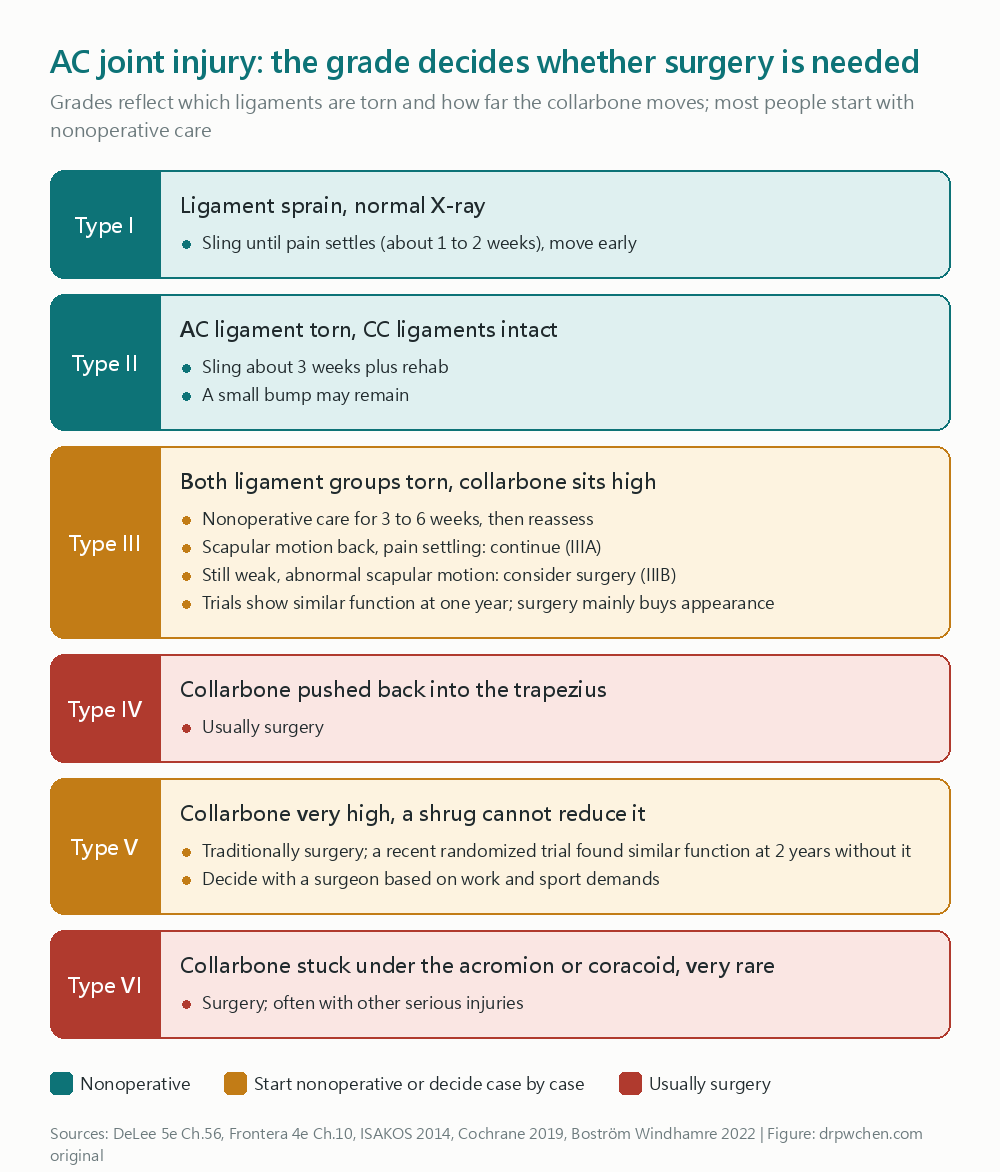
Doctors classify AC joint injuries into six types:
From mild to severe
- Type 1: Ligament sprain, X-rays look normal.
- Type 2: The ligaments next to the joint are torn, but the suspensory ligaments underneath are still intact. The collarbone is slightly elevated.
- Type 3: Both sets of ligaments are torn, and the collarbone is obviously sticking up. This is the type where we debate most often about “whether to operate.”
- Type 4: The collarbone is pushed back into the muscles of the back.
- Type 5: The collarbone is pushed up very high, tearing even the muscle attachments around the shoulder.
- Type 6: The collarbone gets stuck under the bones below the shoulder. Very rare.
Do I need an X-ray? What other tests do I need?
Yes. After a shoulder fall, the most important reason for an X-ray is to rule out fractures. For example, an outer collarbone fracture looks a lot like an AC joint injury from the outside, but we treat it differently.
Doctors usually ask the radiographer to shoot both shoulders together. We need to compare left and right to see the difference. Ultrasound can also look at this joint; it’s even more sensitive than X-rays for mild sprains, and we can compare it to the uninjured side right on the spot.
The old “stress views,” where you held heavy weights during the X-ray, are mostly unnecessary now. Taking that extra shot hurts a lot and doesn’t really help us decide how to treat you.
If I don’t get surgery, how do I recover? How long do I wear the sling?
Type 1, Type 2, and most Type 3s start without surgery:
- You can take the sling off once it stops hurting. About one to two weeks for mild cases, and around three weeks for Type 2.
- Ice it for pain, and start moving it gently within a pain-free range during the first week.
- Early on, avoid “raising your arm out to the side” and “reaching across your chest”—those two motions stress the joint the most.
- If you lift weights, bench presses and overhead presses need to wait a bit longer before you get back to them.
Then rehab will step-by-step add strength training, especially for the muscles that stabilize the shoulder blade. The whole rehab process takes about six to twelve weeks.
That bump on your shoulder? If you don’t have surgery, it’ll probably stay there. The good news is, for most people, it’s mostly a cosmetic issue. It doesn’t really affect strength or movement.
The collarbone is sticking up really high. Do I absolutely need surgery?
Not necessarily. This is the most frequently asked—and most debated—question about AC joint injuries.
Type 3: The current approach is to try conservative treatment for three to six weeks first, then have a doctor re-evaluate it.
- If your shoulder blade movement recovers and the pain slowly decreases, you keep going with conservative care.
- If it’s still very weak after this period, and your shoulder blade moves weirdly, then you talk to an orthopedic surgeon about an operation.
Multiple randomized controlled trials split Type 3 patients into surgery and no-surgery groups. A year later, shoulder function was about the same in both groups. The people who didn’t get surgery actually recovered faster. The main benefits of surgery are that it looks flatter and the X-ray looks more perfectly aligned.
Studies have also found that people who try conservative care first and later switch to surgery get similar results to those who did well with conservative care from the start. So it usually doesn’t hurt to try the conservative route first.
Type 5: In the past, these almost always went to surgery. But a recent randomized trial found that Type 5 patients without surgery had similar function two years later compared to those who had surgery. So now you can discuss it with your doctor and decide based on your work and sports needs.
Types 4 and 6: These usually require surgery.
If you are a heavy manual laborer, a pitcher, or an athlete who does a lot of overhead motions, or if you care a lot about the cosmetic appearance, you might want to discuss the pros and cons of surgery with an orthopedic surgeon earlier.
Can the top of the shoulder hurt even if I didn’t fall?
Yes. The AC joint doesn’t just get injured from falls; it can also be “worn out”:
- People who frequently lift weights: Repetitive bench presses, push-ups, and dips can slowly cause the bone at the end of the collarbone to be resorbed. This is called “distal clavicle osteolysis,” or sometimes “weightlifter’s shoulder.” Modifying your lifting routine and pausing painful movements for at least six weeks usually improves it.
- People past middle age: The AC joint naturally degenerates with age, often showing up alongside other shoulder problems (like the rotator cuff). You can take anti-inflammatories, get injections, and do rehab. Only if those don’t work well would we consider cutting off a small piece of the collarbone end.
Weightlifters can make these adjustments:
- Drop the weight first.
- Don’t grip too wide on the bench press, and stop about 4 to 6 cm above your chest when lowering the bar—don’t let it touch your chest.
- Seen from the side, keep your elbows level with your torso or in front of it at the start and throughout the lift, not dropping behind it.
If an older adult’s X-ray shows a “widened” joint, is it from the recent fall?
Not necessarily. The AC joint starts degenerating when we’re young. By our fifties, the joint space is often already narrowed and has bone spurs.
So when an older adult falls and the X-ray shows the “joint looks weird,” we can’t jump straight to assuming it was from this fall. Doctors will:
- X-ray or scan the other uninjured shoulder to compare.
- Look for signs that only show up with a fresh injury, like it hurting a lot when pressed right on that spot, or fluid building up in the joint.
- Ask if the shoulder had hurt or been injured in the past.
If an older adult can’t lift their arm after a fall, we also have to remember that other parts of the shoulder, like the rotator cuff, might be injured at the same time. We shouldn’t just stare at the AC joint.
When should I rush back to the doctor?
- Your arm feels numb, weak, or your hand’s color or temperature seems off.
- The skin over the collarbone bump is stretched really tight, or there’s a new swollen lump on the back of your shoulder.
- The pain is much worse than expected, or it keeps getting worse after a few days.
- After a few weeks of conservative care, you still can’t lift your arm and it feels very weak.
- You didn’t injure yourself, but the tops of both shoulders hurt, or you have a fever with it.
Further reading: Rotator cuff injury, Shoulder impingement, Shoulder dislocation, Pain at the inner end of the collarbone (sternoclavicular joint)
Below is the exact note from my vault, with only the internal links and copyrighted textbook images removed. The fact-checking workflow and tools are explained at the end.
Acromioclavicular joint injury
Clinical Pearls
- Mechanism: usually falling or taking a direct hit to the acromion with the arm adducted; AC lig. handles horizontal stability, CC lig. (conoid + trapezoid) handles vertical stability
DeLee 5e Ch.56- Rockwood I–VI: I, II conservative; IV, VI surgery; III conservative for 3–6 weeks first, then split into IIIA/IIIB
DeLee 5e Ch.56,Beitzel 2014- Type III multiple RCTs (Cochrane pooled acute AC dislocations without separating types): Surgery does not beat conservative functionally at one year, and conservative recovery is faster; surgery trades for cosmesis and radiographic reduction
Tamaoki 2019,Xie 2024- Type V is no longer an “absolute surgical indication”: In a hook plate RCT, Type V conservative at 24 months had a Constant score of 90 vs 91
Boström Windhamre 2022- Older adults with AC imaging abnormalities should make you think of pre-existing degeneration first: Degeneration starts young, classically showing narrowing + osteophytes; widening means looking for ligament injury or distal clavicle resorption, always compare to the unaffected side
Levangie 5e Ch.7,Martinoli Ch.6
Abbreviations
| Abbreviation | Full Name |
|---|---|
| AC | acromioclavicular |
| CC | coracoclavicular |
| CA | coracoacromial |
| DCO | distal clavicle osteolysis |
| ISAKOS | International Society of Arthroscopy, Knee Surgery and Orthopaedic Sports Medicine |
| ESSKA | European Society of Sports Traumatology, Knee Surgery and Arthroscopy |
| CS | Constant(-Murley) score |
| DASH | Disabilities of the Arm, Shoulder and Hand |
| CCD | coracoclavicular distance |
| RCT | randomized controlled trial |
Disease Background
Definition
- AC joint = the diarthrodial joint between the lateral end of the clavicle and the medial acromion, enveloped by a fibrous capsule
Frontera 4e Ch.10 - The joint contains a meniscus-like disc that degenerates with age, largely nonfunctional after age 40
DeLee 5e Ch.56 - Three sets of ligaments
DeLee 5e Ch.56,Frontera 4e Ch.10- AcromioClavicular lig.: Primary restraint to anteroposterior (horizontal) translation
- CoracoClavicular lig.: conoid (medial, stronger) + trapezoid (lateral); primarily restrains superior-inferior (vertical) translation, “suspends” the scapula from the clavicle
- CoracoAcromial lig.: Does not tear in AC injuries; acts as a secondary restraint against superior migration of the humeral head in massive cuff tears
- Displacement amount dictates which ligament dominates: With small displacements, the AC lig. blocks posterior and superior translation; with large displacements, the conoid becomes the main restraint against superior translation
DeLee 5e Ch.56 - Movement: During 180° of arm elevation, the AC joint itself only moves 5–8°; the 40–50° rotation along the clavicle’s long axis primarily relies on synchronous scapular movement
DeLee 5e Ch.56 - This note covers three clinical entities:
- Traumatic AC separation (Rockwood I–VI)
- Distal clavicle osteolysis (DCO) (weightlifter’s shoulder) → See Distal Clavicle Osteolysis (DCO)
- AC joint osteoarthritis (AC OA) → See AC Joint Osteoarthritis (AC OA)
Epidemiology
- Accounts for about 9% of all shoulder injuries; mostly young men, incomplete types outnumber complete types
DeLee 5e Ch.56 - NCAA data: Mostly player-to-player contact; in sports played by both sexes, men are affected about 5 times more often
Frontera 4e Ch.10 - Traumatic AC injuries are concentrated in the first three decades (before age 30), involving contact sports or falling on an adducted arm
Levangie 5e Ch.7 - Degenerative changes are common from the second decade (teens) on, and by the sixth decade (50s), the joint space is often already narrowed
Levangie 5e Ch.7 - DCO commonly affects weightlifters and heavy manual laborers
Kahn Ch.16,Cahill 1982
Mechanism of Injury
Traumatic Separation
- Direct trauma: Falling or taking a direct hit to the acromion with the arm adducted, pushing the scapula downward while the SC joint keeps the clavicle in place
DeLee 5e Ch.56,Bahr IOC Ch.7 - Indirect trauma: Falling on an outstretched, adducted arm, driving the humeral head superiorly into the acromion
DeLee 5e Ch.56 - Sequence of injury: AC lig. → CC lig. → deltoid and trapezius fascia
DeLee 5e Ch.56 - Direct blow from the side: Compresses the AC joint, injuring the articular surface and disc (the source of future OA/DCO)
Bahr IOC Ch.7
Mechanism of DCO
- Repetitive microtrauma → microfractures in the subchondral bone of the distal clavicle → hyperemic resorption during the repair process (Cahill’s theory, the most accepted)
Kahn Ch.16,Kelley 11e Ch.49 - Divided into two types
Kahn Ch.16,Kelley 11e Ch.49- Atraumatic (more common): Repetitive loading from weightlifting, gymnastics, swimming, etc.
- Post-traumatic: Distal clavicle resorption appears about 4 weeks after an AC trauma
- Alternative theory: Autonomic dysfunction increases clavicular blood flow, leading to bone resorption
Kelley 11e Ch.49
Mechanism of AC OA
- Age-related degenerative process, the disc degenerates first
Kelley 11e Ch.49,DeLee 5e Ch.56 - Post-traumatic: Instability or damage to the articular surface/disc → secondary OA or DCO
Bahr IOC Ch.7 - Weightlifting, repetitive overhead or throwing activities
DeLee 5e Ch.56
Complications
- After conservative treatment: Late arthrosis, persistent instability, DCO
DeLee 5e Ch.56 - Cosmesis: Conservative management will leave a bump on the shoulder; it’s unavoidable without surgery
Frontera 4e Ch.10 - AC pain caused by chronic instability is the most common complication
Frontera 4e Ch.10 - Concomitant fractures: Coracoid fractures are most commonly associated with Type III, via avulsion from the short head of the biceps and coracobrachialis
Martinoli Ch.6 - Heterotopic ossification: Ossification of the CC lig. is usually painless
Frontera 4e Ch.10 - Long-term OA prevalence (systematic review of 3,812 patients, 2–24 years follow-up)
Vossen 2024- Ranged from 6.7% (CC fixation) to 29.3% (conservative) across treatment groups, and increased over time
- No difference in OA prevalence between the injured and uninjured sides. Author’s conclusion: Fear of OA should not dictate whether to operate.
- Included studies leaned heavily surgical (3483 vs 329), most followed for <10 years, variable quality
- Surgical complications → See Surgical Management
Prognosis
- Type I, II: Most are asymptomatic within 3 weeks
Frontera 4e Ch.10- But long-term isn’t perfectly asymptomatic: At a mean follow-up of 10.2 years, 52% have occasional AC symptoms, Constant score 70.5 vs 86.8 on the normal side
Mikek 2008 - Evidence: Low — 23-patient case series, uncontrolled
- About 1/5 have ROM restrictions, but <10% report it affects daily life or sports
Frontera 4e Ch.10
- But long-term isn’t perfectly asymptomatic: At a mean follow-up of 10.2 years, 52% have occasional AC symptoms, Constant score 70.5 vs 86.8 on the normal side
- Type III: Over 70% successfully complete conservative care, with good outcomes at a mean follow-up of 3.5 years
DeLee 5e Ch.56- Those failing conservative care who undergo delayed reconstruction score similarly to those with successful conservative care
DeLee 5e Ch.56 - However, the proportion returning to pre-injury sports levels is lower for those without surgery
DeLee 5e Ch.56
- Those failing conservative care who undergo delayed reconstruction score similarly to those with successful conservative care
- Bahr: The natural history of I–III is generally favorable, and persistent deformity is mostly just a cosmetic issue
Bahr IOC Ch.7
Clinical Assessment
Diagnostic Criteria
- Clinical diagnosis; post-traumatic AC pathology triad: AC point tenderness + AC pain on cross-arm adduction + relief after local anesthetic injection
DeLee 5e Ch.56 - Classification relies on X-ray (Rockwood is fundamentally a radiographic classification); IIIA/IIIB adds functional assessment
DeLee 5e Ch.56,Beitzel 2014 - Diagnostic local injection: ≥50% pain reduction after AC injection is the reference standard in the Walton study
Walton 2004
Evaluation Tools
History
- Fall or blow to the acromion; pain is over the anterosuperior shoulder, and can radiate to the base of the neck, trapezius, and deltoid
Frontera 4e Ch.10 - Pain distribution helps differentiate the source
DeLee 5e Ch.56- AC irritation: The AC joint itself, anterolateral neck, anterolateral deltoid
- Subacromial irritation: Lateral acromion and deltoid, does not radiate to the neck and trapezius
- Provocative motions: Reaching across the chest (reaching into a pocket), reaching behind the back (tucking in a shirt), side-sleeping on the affected side
Frontera 4e Ch.10 - DCO: Insidious AC pain, hurts during bench press, push-ups, dips; relieved by stopping the exercises
Kahn Ch.16,Kelley 11e Ch.49- Adduction movements like a golf swing or buckling a seatbelt will hurt
Kelley 11e Ch.49
- Adduction movements like a golf swing or buckling a seatbelt will hurt
Physical Examination
- Look while sitting or standing: Arm weight makes the clavicular bump more obvious; check both the clavicle and AC bilaterally (can be bilateral injuries)
DeLee 5e Ch.56 - Pain more severe than expected → think coracoid fracture or Type IV (clavicle piercing the trapezius fascia)
DeLee 5e Ch.56 - Shrug test: Shrugging reduces the joint = Type III (deltotrapezial fascia intact); does not reduce = Type V
DeLee 5e Ch.56 - Scapular dyskinesis: Can be reliably evaluated within 10 days of injury, checking if the medial borders of the scapulae are asymmetrical
DeLee 5e Ch.56 - Acute Type III is often too painful to examine properly → re-evaluate at 3–6 weeks
DeLee 5e Ch.56 - Special tests (mostly for AC arthrosis/DCO, less specific for instability types)
DeLee 5e Ch.56- Cross-body adduction: Arm forward flexed to 90° and pulled across the chest; only positive if the pain is at the AC joint itself
DeLee 5e Ch.56 - Active compression (O’Brien): Forward flexion 90°, adduction 10–15°, apply downward pressure in internal rotation; pain at the shoulder top = AC, pain deep anteriorly = labrum
DeLee 5e Ch.56 - AC resisted extension: Resisted extension from the 90° flexed position
DeLee 5e Ch.56 - Paxinos: Thumb pushes the acromion from posterolaterally while the fingers press the mid-clavicle
Walton 2004
- Cross-body adduction: Arm forward flexed to 90° and pulled across the chest; only positive if the pain is at the AC joint itself
- Test accuracy (retrospective on 35 patients diagnosed post-op)
Chronopoulos 2004- Sensitivity: cross-body 77%, resisted extension 72%, active compression 41%
- Specificity: active compression 95%
- Single test PPVs all <30%, NPVs all >94% → good for ruling out; combinations are more useful
- Test accuracy (injection response as reference standard, 38 patients)
Walton 2004- AC tenderness sensitivity 96%, Paxinos 79%, but specificity is low
- Paxinos + bone scan both positive → post-test probability 99%; both negative → LR 0.03, virtually ruled out
Radiography
- Trauma requires a complete shoulder series: Concomitant GH injuries are common
DeLee 5e Ch.56 - Zanca view: Beam tilted 10–15° cephalad, penetration reduced to 50% of a standard AP shoulder, compared on the same film bilaterally
DeLee 5e Ch.56,Kelley 11e Ch.49 - Normal values (varies by book, base it on the uninjured side)
- AC space: 1–3 mm and narrows with age (Zanca)
DeLee 5e Ch.56; 0.3–0.8 cmMcKinnis 5e Ch.15 - CC distance: 1.1–1.3 cm
DeLee 5e Ch.56; 1.0–1.3 cmMcKinnis 5e Ch.15 - CC distance increased by 25–50% compared to the normal side suggests complete CC lig. rupture
DeLee 5e Ch.56
- AC space: 1–3 mm and narrows with age (Zanca)
- Axillary view: Looks for posterior clavicle displacement in Type IV, but recent studies suggest it is hard to interpret
DeLee 5e Ch.56 - Cross-body adduction view (Basamania): Clavicle overriding the acromion = horizontal instability, used for IIIB
DeLee 5e Ch.56,Beitzel 2014 - Stress view
- Method: Bilateral AP taken without weights first, then a stress view with 5–7 kg strapped to each wrist
McKinnis 5e Ch.15 - Currently not recommended routinely: Differentiating between Type II and III has little clinical meaning; it just adds pain without adding accuracy
DeLee 5e Ch.56,Frontera 4e Ch.10 - ESSKA consensus: True AP or bilateral Zanca is sufficient for diagnosis
Rosso 2020
- Method: Bilateral AP taken without weights first, then a stress view with 5–7 kg strapped to each wrist
- Stryker notch view: AC dislocation but normal CC distance → suspect a coracoid fracture
DeLee 5e Ch.56 - DCO: Loss of subchondral bone detail at the distal clavicle, microcystic changes, osteopenia in the lateral 1/3; overt AC widening in late stages
Kelley 11e Ch.49 - AC OA: Subchondral microcysts, sclerosis, osteophytes, joint space narrowing
Kelley 11e Ch.49- Imaging does not always correlate with symptoms: AC degeneration is an age-related change (DePalma)
Kelley 11e Ch.49
- Imaging does not always correlate with symptoms: AC degeneration is an age-related change (DePalma)
Ultrasound
- AC injuries are often mistaken for post-traumatic cuff pathology (closely positioned to the supraspinatus)
Martinoli Ch.6 - For low-grade sprains, ultrasound is more sensitive than X-ray: Can see joint space widening, hematoma or effusion, and bulging of the superior capsule
Martinoli Ch.6 - The CC lig. is obscured by the clavicle and can’t be seen directly; hematoma between the clavicle and coracoid is an indirect sign
Martinoli Ch.6 - Severe types may show tears and hematomas at the deltoid or trapezius attachments
Martinoli Ch.6 - Measurement: Measure the AC width in the coronal plane and compare it to the unaffected side; normal width ≤6 mm
Martinoli Ch.6- AC Index = unaffected side width / affected side width; Tossy I is ~1.0, II is ~0.5, III is ~0.2
Kock 1996 - Index below 0.3 corresponds to Tossy III
Kock 1996 - ⚠️ Martinoli’s original text flipped the numerator and denominator (affected/unaffected), contradicting the very values it cited; go by the original study.
- AC Index = unaffected side width / affected side width; Tossy I is ~1.0, II is ~0.5, III is ~0.2
- Post-traumatic DCO: Irregular cortical erosions at the clavicle end, joint widening, and effusion; the acromial end is normal
Martinoli Ch.6 - AC OA: Joint space narrowing, osteophytes, para-articular cysts
Martinoli Ch.6 - With massive cuff tears, a cyst can pop up over the AC joint (Geyser sign)
Martinoli Ch.6
MRI
- Among symptomatic AC joints, 80% show reactive bone edema (distal clavicle, acromion, or both), versus 0% in asymptomatic controls
Shubin Stein 2006- Bone edema predicts symptoms better than the degree of degeneration
- DCO: high T2 signal in the distal clavicle
Kahn Ch.16 - Bone scan: increased distal clavicle uptake in early DCO, before X-ray changes appear, but not specific
Kahn Ch.16,Kelley 11e Ch.49
AC Widening in Older Adults: Distinguishing Acute from Chronic
- AC trauma usually occurs before age 30; degeneration starts in the teens, and by the 50s the space is often narrowed
Levangie 5e Ch.7 - Normal aging involves narrowing (normal Zanca 1–3 mm decreases with age), so widening itself is not normal aging
DeLee 5e Ch.56 - Non-traumatic sources of widening: Pseudowidening caused by distal clavicle resorption
Martinoli Ch.6,Kelley 11e Ch.49- RA, DCO, hyperparathyroidism, infection
- Supporting evidence to look for in trauma: Acute point tenderness, bulging superior capsule and effusion, relative superior clavicular displacement, increased CC distance
Martinoli Ch.6,DeLee 5e Ch.56 - Practice: Film or scan the uninjured side to compare, and judge based on history and X-ray osteophytes/cysts to see if it’s old
Bahr IOC Ch.7,Martinoli Ch.6 - Symptomatic AC degeneration in those ≥50 years old is 86% associated with cuff pathology (about half are full-thickness tears)
Brown 2000- This is the group where AC degeneration requires surgery, not the trauma group; Frontera misread this as trauma with concomitant injury.
Classification
Rockwood (Tossy I–III expanded to I–VI) + ISAKOS Modification
| Type | AC lig. | CC lig. | Deltotrapezial fascia | Imaging / Clinical | Default Management |
|---|---|---|---|---|---|
| I | Sprained | Intact | Intact | Normal X-ray, AC point tenderness only | Conservative |
| II | Torn | Sprained but intact | Intact | Mild superior displacement of the clavicle tip, horizontal instability | Conservative |
| IIIA (stable) | Torn | Torn | Mildly injured | 100% superior displacement; no overriding on cross-body view; obvious improvement after 3–6 weeks of conservative care | Conservative |
| IIIB (unstable) | Torn | Torn | Mildly injured | Clavicle overriding the acromion on cross-body view; persistent scapular dyskinesis, weakness, poor ROM after 3–6 weeks of conservative care | Consider surgery |
| IV | Torn | Torn | Injured (clavicle posteriorly displaced into the trapezius) | Clavicle displaced posteriorly on axillary view; skin tenting, posterior fullness | Surgery |
| V | Torn | Torn | Stripped from the clavicle | CC distance increased by 100–300%; irreducible on shrug test | Traditional surgery, evidence is weakening (see below) |
| VI | Torn | Torn | Potentially injured | Clavicle lodged subacromial or subcoracoid; often accompanied by severe trauma and transient paresthesia | Surgery |
- Table is based on
DeLee 5e Ch.56Table 56.2,Frontera 4e Ch.10Table 10.1,Beitzel 2014 - Tossy 1963 was I–III, Rockwood 1984 expanded to VI; purely a radiographic classification
DeLee 5e Ch.56 - IIIA/IIIB is a functional rather than anatomical reclassification (ISAKOS 2014)
Beitzel 2014 - ESSKA consensus: ISAKOS modified Rockwood is valid; the acute vs. chronic cutoff is 3 weeks
Rosso 2020 - Displacement cutoffs vary across textbooks, don’t memorize a single number
- Type II: DeLee says 50% vertical subluxation
DeLee 5e Ch.56; McKinnis says AC space 1.0–1.5 cm, CC increased by 25–50%McKinnis 5e Ch.15 - Type III: DeLee says 100% superior displacement
DeLee 5e Ch.56; Frontera says CC distance increased by 25–100%Frontera 4e Ch.10
- Type II: DeLee says 50% vertical subluxation
DCO Classification
- Atraumatic vs. post-traumatic
Kahn Ch.16 - Post-traumatic DCO is self-limiting: Appears weeks to years after injury, gradually repairs within 4–6 months
Martinoli Ch.6 - Changes are only at the clavicular end, the acromion is normal
Martinoli Ch.6
Differential Diagnosis
| Condition | Key Differentiator | Source |
|---|---|---|
| Lateral clavicle fracture | Looks like Type II/III externally if fracture is medial to CC lig.; pain is over the clavicle; X-ray is a must | Frontera 4e Ch.10, AAOS Clavicle Fx 2022 |
| Coracoid fracture | AC dislocated but normal CC distance; Stryker notch view | DeLee 5e Ch.56, Martinoli Ch.6 |
| Rotator cuff pathology / impingement | Pain is in the middle of the arc of motion, subsides afterward; resisted cuff testing hurts with arm at the side | Frontera 4e Ch.10 |
| SLAP / labral tear | O’Brien test hurts deep anteriorly, relieved in external rotation | DeLee 5e Ch.56, Kahn Ch.16 |
| DCO | No acute trauma; history of weightlifting; X-ray shows distal clavicular resorption | Kahn Ch.16 |
| AC OA | Older age, joint space narrowing, osteophytes; symptoms and imaging may not correlate | Kelley 11e Ch.49 |
| Septic AC | Joint space widening, bony erosions, mobile debris | Martinoli Ch.6 |
| RA/hyperPTH/sarcoidosis/Gorham disease | Think systemic causes if distal clavicular resorption is bilateral | Kelley 11e Ch.49, Kahn Ch.16 |
| Cervical radiculopathy | Abnormal neck and neurological exam | Frontera 4e Ch.10 |
| Os acromiale | Unfused ossification center seen on ultrasound or axillary view | Frontera 4e Ch.10, Martinoli Ch.6 |
Red Flags and Referral
- Nerve or vascular injury → Indicates a force greater than a simple AC injury
Frontera 4e Ch.10 - Type IV/V/VI or skin tenting → Early referral to orthopedics
Frontera 4e Ch.10,DeLee 5e Ch.56 - Clavicle fracture medial to the CC lig. (risk of displacement, delayed union or nonunion) → Referral to orthopedics
Frontera 4e Ch.10- Displaced lateral clavicle fracture with torn CC lig.: AAOS workgroup feels this may benefit from surgery (Consensus)
AAOS Clavicle Fx 2022
- Displaced lateral clavicle fracture with torn CC lig.: AAOS workgroup feels this may benefit from surgery (Consensus)
- Type III in a throwing athlete, or unclassifiable injuries → Orthopedic evaluation
Bahr IOC Ch.7 - Bilateral DCO → Workup for RA, hyperparathyroidism, sarcoidosis
Kelley 11e Ch.49 - AC widening plus bony erosions and fever → Think infection
Martinoli Ch.6
Clinical Management
Management Algorithm
Algorithm
- Decision axis = Rockwood classification × function after 3–6 weeks conservative care × patient needs
DeLee 5e Ch.56- Rule out first: Fractures (lateral clavicle, coracoid), neurovascular injury → Orthopedic referral
Frontera 4e Ch.10- Type I, II: Conservative (sling until pain-free, early ROM, 4-phase rehab)
DeLee 5e Ch.56- Type III
- Always try conservative care first for 3–6 weeks (ISAKOS)
DeLee 5e Ch.56- Re-evaluate at 3–6 weeks, finalize decision by 3 months at the latest: IIIA → continue conservative; IIIB (persistent scapular dyskinesis + overriding on cross-body view) → consider surgery
DeLee 5e Ch.56,Beitzel 2014- Weigh toward surgery: Heavy laborers, pitchers or quarterbacks, those able to comply with post-op protocols
DeLee 5e Ch.56- Athletes in-season: Can return to play after intra-articular lidocaine injection; off-season, functional rehab for up to 3 months before deciding
DeLee 5e Ch.56- Type IV, VI: Surgery; can try closed reduction to Type III first then manage conservatively
DeLee 5e Ch.56- Type V: Traditionally surgery, but RCT shows conservative function is not inferior at 24 months → Discuss with the patient
DeLee 5e Ch.56,Boström Windhamre 2022- Surgical timing: Chronic (>3 weeks) requires biologic graft augmentation
DeLee 5e Ch.56,Rosso 2020- DCO: Stop or modify workout for ≥6 weeks + NSAIDs + injection if needed → Distal clavicle excision if this fails
Kahn Ch.16- AC OA: NSAIDs, injection, rehab, avoid provocative motions → Distal clavicle excision if this fails
Kelley 11e Ch.49,Bahr IOC Ch.7
Treatment Goals
- Pain-free, full ROM, normal strength, unrestricted activity, whether surgical or not
DeLee 5e Ch.56 - Determined by patient demands and expectations for the shoulder (occupation, sports, season timing, concern for cosmesis)
DeLee 5e Ch.56
Conservative Management
Acute Phase
- Sling, ice, analgesics; short-term immobilization for 3–7 days
DeLee 5e Ch.56 - Sling until pain-free: ~1–2 weeks for Type I, ~3 weeks for Type II
DeLee 5e Ch.56- Frontera says 1–6 weeks, average 2–3 weeks
Frontera 4e Ch.10
- Frontera says 1–6 weeks, average 2–3 weeks
- Relative rest; avoid complete immobilization if possible
Frontera 4e Ch.10 - Ice massage for 5–10 minutes or cold pack for 20 minutes, repeatable every 2 hours
Frontera 4e Ch.10 - Start ROM within the first week post-injury
DeLee 5e Ch.56
Rehabilitation (4 phases, Gladstone)
- Phase 1: Pain control, protected ROM, isometrics, closed kinetic chain scapular exercises
DeLee 5e Ch.56- Active-assisted ROM in the scapular plane (abduction 30–45°, forward flexion 30–40°) up to the onset of pain
- Avoid abduction/elevation: Clavicle upward rotation tensions the AC lig.
- Progression criteria: 75% ROM, AC nearly non-tender, anterior/middle deltoid and upper trapezius MMT 4/5
- Phase 2: Progression to full painless ROM, isotonic strength
DeLee 5e Ch.56- Bench press and military press temporarily restricted (increases AC stress)
- Progression criteria: Painless ROM, non-tender, strength 75% of the contralateral side
- Phase 3: Full shoulder complex strength and endurance (dumbbell front raises, abduction, shrugs, bench press)
DeLee 5e Ch.56 - Phase 4: Sports-specific; criteria = full ROM, pain-free, isokinetic strength approaching 100% of the contralateral side
DeLee 5e Ch.56 - 6–12 weeks to complete the full rehab protocol
DeLee 5e Ch.56 - Type III should be referred to PT early to restore mobility and scapular control
DeLee 5e Ch.56 - Early strengthening: 1–5 lb dumbbells or resistance bands for internal/external rotation, 10–20 reps daily
Frontera 4e Ch.10
Injection
- Diagnostic: Complete pain relief after local anesthetic injection confirms Type I
Frontera 4e Ch.10 - Type I or mild II: If full ROM and symmetric strength return post-injection, can return to play or work the same day
Frontera 4e Ch.10- Cannot use injections to substitute for relative rest in higher-grade injuries; avoid provocative motions for 1 week post-injection
Frontera 4e Ch.10
- Cannot use injections to substitute for relative rest in higher-grade injuries; avoid provocative motions for 1 week post-injection
- Accuracy: Blind injections truly enter the joint 40–60% of the time
Frontera 4e Ch.10- Cadaver study: Ultrasound-guided 10/10 in joint vs. palpation 4/10
Peck 2010 - It remains unclear whether guidance improves clinical outcomes
Frontera 4e Ch.10
- Cadaver study: Ultrasound-guided 10/10 in joint vs. palpation 4/10
- A needle that is too long might enter the subacromial space, causing diagnostic confusion
Frontera 4e Ch.10
Type III: Evidence for Surgery vs. Conservative Care
- Arguments for a conservative-first approach
- Cochrane (acute AC dislocations, types not separated; 5 RCTs + 1 quasi-RCT, 357 patients; no subgroup analysis by type possible)
Tamaoki 2019- One-year DASH no difference (2 trials, 112 patients; MD 0.73, 95% CI -2.70 to 4.16)
- Conservative group better function at 6 weeks (faster recovery)
- Treatment failure requiring reoperation: 14/168 vs 15/174, RR 0.99
- More adverse events in the surgery group (RR 2.82), though mostly from now-rarely-used hardware (like K-wires)
- Evidence: Low to very low — unblinded, small sample sizes
- Type III specific RCT meta-analysis (4 studies, 244 patients): Long-term CS no difference (MD 4.82, 95% CI -6.42 to 16.06)
Xie 2024- Surgery: Better early pain relief, better CCD
- Surgery: More post-traumatic OA and hardware issues
- III–V RCT meta-analysis (367 patients): No difference in function or reoperation; higher complication rate in surgery group (no difference in the Type III subgroup)
Wen 2026 - Early large synthesis: 88% surgical vs. 87% conservative satisfaction
Phillips 1998- Reoperation 59% vs 6%, infection 6% vs 1%, deformity 3% vs 37%
- Reoperations included hardware removal; data mostly from older techniques
- Delayed reconstruction outcomes are similar to successful conservative care → Trying conservative first does no harm
DeLee 5e Ch.56 - Survey: 86% of AOSSM members prefer conservative; 69% of MLB team physicians
DeLee 5e Ch.56
- Cochrane (acute AC dislocations, types not separated; 5 RCTs + 1 quasi-RCT, 357 patients; no subgroup analysis by type possible)
- Arguments supporting surgery (must be stated honestly)
- RCT (Type III, Weaver-Dunn, 70 randomized, 60 completed)
Lara 2026- 24-month DASH no difference
- But UCLA shoulder score was higher, cosmetic dissatisfaction 14.8% vs 44% (both favoring surgery)
- Meta-analysis (19 studies, 954 patients, III–V)
Chang 2018- Surgery: Better cosmesis (OR 0.05) and radiographic reduction; CS 3.14 points better but possibly not clinically significant
- Conservative: Faster return to work, fewer hardware complications and infections
- ACORN RCT (III–IV, ages 16–35, 60 patients): No functional difference at 1 year
Murray 2018- But a substantial proportion of conservative patients were dissatisfied, 5 crossed over to delayed surgery; could not identify factors predicting failure
- Systematic review: Surgery group seems to have less persistent pain, but the difference was not statistically significant; insufficient evidence for functional conclusions
Longo 2017
- RCT (Type III, Weaver-Dunn, 70 randomized, 60 completed)
- Overall interpretation
- For most people: Start conservatively, then triage by IIIA/IIIB at 3–6 weeks
DeLee 5e Ch.56,Beitzel 2014 - Surgery trades for cosmesis, radiographic reduction, and some subjective scores; the cost is hardware and reoperation risks
- Average patient age in these studies is ~30–54; evidence is still sparse for high-demand athletes and for ages outside this range (e.g., older adults)
Wen 2026
- For most people: Start conservatively, then triage by IIIA/IIIB at 3–6 weeks
Type V: The Old Consensus is Weakening
- Textbooks: IV–VI generally go to surgery
DeLee 5e Ch.56,Frontera 4e Ch.10 - RCT (hook plate vs. physical therapy, stratified for III and V, 124 patients)
Boström Windhamre 2022- 3 months: Conservative group better; 6, 12, 24 months: no difference
- 24-month CS: Type V conservative 90 vs. surgery 91
- Conservative group: 18% (types III and V combined, 11 patients; 5 type V) crossed over to surgery within 19 months
- Only compared against hook plates, cannot be generalized to modern anatomic reconstructions
- Case series (22 patients, average age 42): 77% returned to work, but functional scores were low; younger patients more likely to have normal scores
Dunphy 2016 - Canadian multicenter RCT (III–V, 83 patients, hook plate): 2-year DASH no difference; conservative better early on, fewer reoperations
COTS 2015 - Meta-analysis: Extremely little evidence for Type IV, limited data for Type V
Wen 2026
Surgical Management
Procedural Overview
- Over 75 techniques described, no gold standard
DeLee 5e Ch.56 - Basic types
DeLee 5e Ch.56- Direct AC fixation (pin, screw, hook plate)
- CA lig. transfer (Weaver-Dunn) ± distal clavicle excision
- Anatomic CC reconstruction (tendon graft, suspensory device)
- Principles
DeLee 5e Ch.56- Chronic (>3 weeks) use biologic augmentation with a tendon graft
- Preference for anatomic reconstruction
- Acute with mild AC arthrosis: can preserve the distal clavicle (excision increases horizontal translation); chronic or with existing arthrosis: can excise
- ESSKA consensus: Acute uses arthroscopically assisted anatomic reconstruction + suspensory device, no biologic augmentation needed; chronic uses a tendon graft to reconstruct CC and AC
Rosso 2020 - Network meta-analysis (26 RCTs, 1581 patients): Procedures adding AC fixation or graft augmentation have better function, CCD, and recurrence rates, at the cost of longer surgical time
Bi 2023- Smaller evidence pointing the same way: in type V treated with double Endobutton, adding AC capsule repair gave better CCD and maintenance of reduction (retrospective, 23 patients, Level III)
Maden 2025
- Smaller evidence pointing the same way: in type V treated with double Endobutton, adding AC capsule repair gave better CCD and maintenance of reduction (retrospective, 23 patients, Level III)
Surgical Complications
- Hook plate/K-wire complication rates are the highest (26.3%); unplanned reoperation across all techniques is 0.9–5.4%
Moatshe 2018 - Anatomic CC reconstruction (27 cases)
Milewski 2012- Coracoid tunnel group 8/10 (80%): Coracoid fracture, loss of reduction, intraoperative fixation failure
- Coracoid loop group 6/17 (35%): Clavicle fracture, loss of reduction, infection, adhesive capsulitis
- The “up to 80%” cited by DeLee is from this 10-patient subgroup, not the overall rate
- Smooth K-wires are essentially abandoned: Can migrate to the lung, spinal canal, or subclavian artery
DeLee 5e Ch.56 - Infection 0–9% (average 6%)
DeLee 5e Ch.56 - Type V postoperative MRI (46 patients, follow-up of at least 24 months): neither hook plate nor Endobutton restored native CC ligament thickness, yet functional scores were good
Koluman 2026- Complication patterns differed: more AC arthritis/osteolysis after hook plate, more implant-related problems after Endobutton
- Loss of reduction and recurrent instability mostly correlate with younger patients failing to comply with immobilization and rehab
DeLee 5e Ch.56 - Post-op AC arthrosis, distal clavicle re-calcification or DCO after excision requiring revision, CC lig. ossification
Frontera 4e Ch.10
Post-operative Rehabilitation
- Distal clavicle excision alone: Immobilize 1–3 days → ROM; begin strengthening at 4–6 weeks; return to sports or lifting at 3 months
DeLee 5e Ch.56- Power athletes often need 6–12 months to return to peak strength
- CC reconstruction (AAOS open anatomic CC reconstruction protocol)
AAOS Postop Rehab 2018 Ch.3- Rationale: A tendon graft in a bone tunnel needs about 12 weeks for solid tendon-to-bone healing
- Phase I (0–6/8 weeks): Platform brace (Lerman, Gunslinger) worn full-time to counteract the downward pull of arm weight
- Phase II (6–12 weeks): Closed kinetic chain ROM, scapular control; caution with internal rotation behind the back, cross-body adduction, and end-range forward flexion
- Phase III (12–18 weeks): Begin isotonic strengthening
- Phase IV (4–6 months): Throwing, swimming, tennis, golf at 4–5 months; contact sports after 6 months
- DeLee’s version: Brace 4–6 weeks for acute repairs, 6–12 weeks for chronic; begin strengthening at 12 weeks; heavy lifting at 3–4.5 months; contact sports at least 6 months out
DeLee 5e Ch.56 - Before return to play: Upper extremity power testing must be within 10–15% of the contralateral side
DeLee 5e Ch.56
Distal Clavicle Osteolysis (DCO)
- Conservative
Kahn Ch.16- Avoid provocative exercises for at least 6 weeks, NSAIDs, ice massage
- Physical therapy usually isn’t helpful unless there’s concomitant shoulder pathology
- Intra-articular steroids: Short-term pain relief and diagnostic confirmation
- Modifying workouts (for those unwilling to stop entirely)
Kahn Ch.16- Decrease weight
- Barbell grip less than 1.5 times bi-acromial width
- During bench press, control the descent and stop 4–6 cm above the chest
- Don’t let elbows drop below the coronal plane at the start and during the movement
- Original study: 46 male athletes (45 weightlifters)
Cahill 1982- 21 underwent distal clavicle excision; the 19 followed up had symptom relief, and most could continue training
- The 25 who didn’t get surgery also improved, but only if they stopped or modified their training and avoided heavy lifting
- Bilateral: One long-term follow-up of atraumatic DCO found 70% were bilateral; patients must be warned early
Kelley 11e Ch.49 - Failure of conservative care or unwillingness to modify training → Orthopedic referral for distal clavicle excision
Kahn Ch.16
AC Joint Osteoarthritis (AC OA)
- Conservative: Heat, NSAIDs, steroid injections, shoulder rehab, avoiding painful postures
Kelley 11e Ch.49- If symptoms persist, can repeat injections every 3 months
Kelley 11e Ch.49 - Complete resolution can take a long time
Kelley 11e Ch.49
- If symptoms persist, can repeat injections every 3 months
- Frequently coexists with subacromial impingement and cuff pathology → If necessary, do staged AC and subacromial injections to find the true pain source
Kelley 11e Ch.49 - Surgery: Distal clavicle excision (Gurd, Mumford)
Kelley 11e Ch.49- Open: Good results, but risks injuring the deltotrapezial fascia or rupturing the anterior deltoid
- Arthroscopic: Similar results, fewer complications
- High rate of concomitant shoulder pathology with symptomatic AC degeneration → The original author recommends concomitant shoulder arthroscopy during AC surgery
Brown 2000
Evidence × Setting Matrix
| Intervention / Setting | Evidence | Conclusion | Primary Source |
|---|---|---|---|
| Type I, II conservative | Consensus; no evidence supports surgery | Standard of care; about half have occasional symptoms at 10 years | DeLee 5e Ch.56, Mikek 2008 |
| Type III surgery vs. conservative | Low (Cochrane); consistent across multiple RCTs | No difference in 1-year function; conservative recovers faster; surgery has better cosmesis and reduction | Tamaoki 2019, Xie 2024, Lara 2026 |
| Type III try conservative 3–6 weeks first then split IIIA/IIIB | Expert consensus (ISAKOS) | Those who fail have similar outcomes with delayed reconstruction | Beitzel 2014, DeLee 5e Ch.56 |
| Type V hook plate vs. conservative | Single RCT | No difference at 24 months; 18% of the conservative group (types III and V combined) crossed over to surgery | Boström Windhamre 2022 |
| Surgical technique selection | Network meta-analysis (RCTs) | Adding AC fixation or graft augmentation is better | Bi 2023 |
| Ultrasound-guided AC injection | Cadaver study | Accuracy 100% vs 40%; clinical outcome difference remains unclear | Peck 2010, Frontera 4e Ch.10 |
| DCO conservative vs. excision | Case series | Stopping or modifying training mostly improves it; excision usually allows return to training | Cahill 1982, Kahn Ch.16 |
How These Notes Were Produced
This note was originally my own reference compiled for clinical and teaching purposes. Before putting it on the site this time, I re-checked the evidence entirely.
Materials Used
- Textbooks in PM&R, orthopedics, and rheumatology: DeLee Orthopaedic Sports Medicine 5e, Frontera Essentials 4e, Kahn, Kelley’s Textbook of Rheumatology 11e, IOC Manual of Sports Injuries, AAOS Postoperative Orthopaedic Rehabilitation, Martinoli Ultrasound, McKinnis Fundamentals of Musculoskeletal Imaging 5e, Levangie Joint Structure and Function 5e
- Society consensuses and guidelines: ISAKOS 2014 Rockwood modification consensus, ESA-ESSKA 2020 Delphi consensus, AAOS 2022 clavicle fracture guidelines (lateral clavicle segment)
- Systematic reviews and meta-analyses: Cochrane 2019, meta-analyses of RCTs for Type III and Types III–V, network meta-analysis of surgical techniques, review of long-term osteoarthritis prevalence
- RCTs: Canadian multicenter trial, ACORN trial, hook plate trial for Types III and V, 2026 Type III trial
- Original research: Two papers on physical exam accuracy, ultrasound AC index, MRI bone edema, the original case series on distal clavicle osteolysis
Tools Used
audit_note.py— A custom note format auditing script I wrote to check citation placements, images, and heading structure- Textbook chapter reverse-lookup script — Ensures every “Book Title + Chapter Number” actually matches that chapter
- PubMed MCP — Found systematic reviews and RCTs, filled in bibliographic details (volume, issue, pages, DOIs, PMIDs), and read abstracts one by one to verify numbers
- textbook_search and local textbook markdown indices — Searched and read the original textbook texts paragraph by paragraph
- OpenEvidence — Did an independent cross-search on evidence for managing Type III and V; every claim in the response was verified against the original text before adoption
- Custom Python/PIL drawing scripts — Generated the classification and management diagrams and the cover (
scripts/figures/) - Hugo’s bilingual workflow
i18n_sync.py— Machine-translated the English version first, then manually proofread the medical terminology
This workflow itself is documented in textbook-to-note. If you also have a Claude or Codex subscription, this kind of mechanical labor of fact-checking and organizing can be delegated to it, leaving the judgment calls to yourself. You can start with Getting started with AI: Installation and first steps and How to talk to AI agents.
About This Version
The professional version is taken directly from my own clinical notes, not rewritten for the blog. It retains the citation style of my notes (Book + Chapter, or Author + Year, with the full bibliography below). Copyrighted textbook and journal images are omitted here; the diagrams are re-drawn by me.
If I’ve misunderstood anything, corrections are welcome.
Reference
Textbooks
- DeLee 5e Ch.56 — Ziegler CG, Laurencin SJ, Frank RM, et al. Injury to the Acromioclavicular and Sternoclavicular Joints. DeLee, Drez & Miller’s Orthopaedic Sports Medicine, 5e, 2020
- Frontera 4e Ch.10 — Rizzo TD Jr. Acromioclavicular Injuries. Frontera: Essentials of Physical Medicine and Rehabilitation, 4e, 2019
- Kahn Ch.16 — Sutaria RG. Acromioclavicular Joint Pathology. Kahn: Musculoskeletal Sports and Spine Disorders, 1e, 2018
- Kelley 11e Ch.49 — Shoulder Pain. Firestein & Kelley’s Textbook of Rheumatology, 11e, 2021
- Bahr IOC Ch.7 — Shoulder. Bahr: The IOC Manual of Sports Injuries, 1e, 2012
- AAOS Postop Rehab 2018 Ch.3 — Acromioclavicular Separations. AAOS: Postoperative Orthopaedic Rehabilitation, 2018
- Martinoli Ch.6 — Bianchi S, Martinoli C. Shoulder, Ultrasound of the Musculoskeletal System, 1e, 2007
- McKinnis 5e Ch.15 — McKinnis LN. Radiologic Evaluation of the Shoulder, Fundamentals of Musculoskeletal Imaging, 5e, 2022
- Levangie 5e Ch.7 — Levangie PK, Norkin CC. The Shoulder Complex, Joint Structure and Function, 5e, 2011
Society Consensus and Guidelines
- Beitzel 2014 — Beitzel K, Mazzocca AD, Bak K, et al. ISAKOS upper extremity committee consensus statement on the need for diversification of the Rockwood classification for acromioclavicular joint injuries. Arthroscopy. 2014;30(2):271-278. doi:10.1016/j.arthro.2013.11.005. PMID: 24485119
- Rosso 2020 — Rosso C, Martetschläger F, Saccomanno MF, et al. High degree of consensus achieved regarding diagnosis and treatment of acromioclavicular joint instability among ESA-ESSKA members. Knee Surg Sports Traumatol Arthrosc. 2021;29(7):2325-2332 (epub 2020). doi:10.1007/s00167-020-06286-w. PMID: 32980887
- AAOS Clavicle Fx 2022 — Management of Clavicle Fractures CPG, American Academy of Orthopaedic Surgeons, 2022 (Lateral clavicle fracture: Consensus)
Journal Articles
- Tamaoki 2019 — Tamaoki MJ, Lenza M, Matsunaga FT, Belloti JC, Matsumoto MH, Faloppa F. Surgical versus conservative interventions for treating acromioclavicular dislocation of the shoulder in adults. Cochrane Database Syst Rev. 2019;10(10):CD007429. doi:10.1002/14651858.CD007429.pub3. PMID: 31604007
- Xie 2024 — Xie C, Fan S, Chen L, Huang L, Chen C, Luo H, et al. Comparative efficacy of operative versus conservative treatment for Rockwood type III acromioclavicular joint dislocation: a systematic review and meta-analysis of randomized controlled trials. BMC Musculoskelet Disord. 2024;25(1):960. doi:10.1186/s12891-024-08100-x. PMID: 39587562
- Wen 2026 — Wen L, Chen C, Liu K, Chen G. Surgical versus conservative treatment of acute Rockwood type III-V acromioclavicular joint dislocation: a systematic review and meta-analysis of randomized controlled trials. Injury. 2026;57(4):113125. doi:10.1016/j.injury.2026.113125. PMID: 41724020
- Lara 2026 — Lara PHS, Lima EBS, Andreoli CV, Pochini AC, et al. Functional, Radiological, and Scapular Motion Evaluation of Surgical Versus Nonsurgical Treatment of Type 3 Acromioclavicular Dislocations: A Randomized Controlled Trial With 24 months’ Follow-up. Am J Sports Med. 2026;54(1):118-127. doi:10.1177/03635465251395220. PMID: 41476398
- Boström Windhamre 2022 — Boström Windhamre H, von Heideken J, Une-Larsson V, Ekström W, et al. No difference in clinical outcome at 2-year follow-up in patients with type III and V acromioclavicular joint dislocation treated with hook plate or physiotherapy: a randomized controlled trial. J Shoulder Elbow Surg. 2022;31(6):1122-1136. doi:10.1016/j.jse.2021.12.003. PMID: 35007749
- Murray 2018 — Murray IR, Robinson PG, Goudie EB, Duckworth AD, et al. Open Reduction and Tunneled Suspensory Device Fixation Compared with Nonoperative Treatment for Type-III and Type-IV Acromioclavicular Joint Dislocations: The ACORN Prospective, Randomized Controlled Trial. J Bone Joint Surg Am. 2018;100(22):1912-1918. doi:10.2106/JBJS.18.00412. PMID: 30480595
- COTS 2015 — Canadian Orthopaedic Trauma Society. Multicenter Randomized Clinical Trial of Nonoperative Versus Operative Treatment of Acute Acromio-Clavicular Joint Dislocation. J Orthop Trauma. 2015;29(11):479-487. doi:10.1097/BOT.0000000000000437. PMID: 26489055
- Chang 2018 — Chang N, Furey A, Kurdin A. Operative Versus Nonoperative Management of Acute High-Grade Acromioclavicular Dislocations: A Systematic Review and Meta-Analysis. J Orthop Trauma. 2018;32(1):1-9. doi:10.1097/BOT.0000000000001004. PMID: 29257778
- Longo 2017 — Longo UG, Ciuffreda M, Rizzello G, et al. Surgical versus conservative management of Type III acromioclavicular dislocation: a systematic review. Br Med Bull. 2017;122(1):31-49. doi:10.1093/bmb/ldx003. PMID: 28334148
- Phillips 1998 — Phillips AM, Smart C, Groom AF. Acromioclavicular dislocation. Conservative or surgical therapy. Clin Orthop Relat Res. 1998;(353):10-17. PMID: 9728155
- Maden 2025 — Maden M, Bacaksiz T, Akan I, Kazimoglu C. Does capsule repair improve the clinical and radiological outcomes of the acute Rockwood type V acromioclavicular dislocations treated with the double endobutton technique? Eur J Trauma Emerg Surg. 2025;51(1):104. doi:10.1007/s00068-025-02786-x. PMID: 39945858
- Koluman 2026 — Koluman AC, Tingir M, Ziroglu N, Ercin E, Moroder P. Incomplete Restoration of Coracoclavicular Ligament Thickness After Surgical Treatment of Rockwood Type 5 Acromioclavicular Dislocations: A Contralateral-Controlled MRI Study With Functional Outcomes. Am J Sports Med. 2026;54(8):2011-2020. doi:10.1177/03635465261448213. PMID: 42227600
- Bi 2023 — Bi AS, Robinson J, Anil U, Hurley ET, et al. Treatment options for acute Rockwood type III-V acromioclavicular dislocations: a network meta-analysis of randomized controlled trials. J Shoulder Elbow Surg. 2023;32(6):1146-1158. doi:10.1016/j.jse.2023.01.039. PMID: 36871607
- Moatshe 2018 — Moatshe G, Kruckeberg BM, Chahla J, et al. Acromioclavicular and Coracoclavicular Ligament Reconstruction for Acromioclavicular Joint Instability: A Systematic Review of Clinical and Radiographic Outcomes. Arthroscopy. 2018;34(6):1979-1995.e8. doi:10.1016/j.arthro.2018.01.016. PMID: 29573931
- Milewski 2012 — Milewski MD, Tompkins M, Giugale JM, Carson EW, Miller MD, Diduch DR. Complications related to anatomic reconstruction of the coracoclavicular ligaments. Am J Sports Med. 2012;40(7):1628-1634. doi:10.1177/0363546512445273. PMID: 22553116
- Dunphy 2016 — Dunphy TR, Damodar D, Heckmann ND, Sivasundaram L, Omid R, Hatch GF. Functional Outcomes of Type V Acromioclavicular Injuries With Nonsurgical Treatment. J Am Acad Orthop Surg. 2016;24(10):728-734. doi:10.5435/JAAOS-D-16-00176. PMID: 27579816
- Vossen 2024 — Vossen RJM, Verstift D, Van Etten-Jamaludin FS, Hamans BJ, van den Bekerom MPJ, Verweij LPE, et al. Choice of Acromioclavicular Dislocation Treatment Should Not Be Influenced by Risk of Development of Acromioclavicular Osteoarthritis: A Systematic Review With Meta-Analysis. JBJS Rev. 2024;12(9). doi:10.2106/JBJS.RVW.24.00085. PMID: 39226400
- Mikek 2008 — Mikek M. Long-term shoulder function after type I and II acromioclavicular joint disruption. Am J Sports Med. 2008;36(11):2147-2150. doi:10.1177/0363546508319047. PMID: 18583520
- Chronopoulos 2004 — Chronopoulos E, Kim TK, Park HB, Ashenbrenner D, McFarland EG. Diagnostic value of physical tests for isolated chronic acromioclavicular lesions. Am J Sports Med. 2004;32(3):655-661. doi:10.1177/0363546503261723. PMID: 15090381
- Walton 2004 — Walton J, Mahajan S, Paxinos A, et al. Diagnostic values of tests for acromioclavicular joint pain. J Bone Joint Surg Am. 2004;86(4):807-812. doi:10.2106/00004623-200404000-00021. PMID: 15069148
- Kock 1996 — Kock HJ, Jurgens C, Hirche H, Hanke J, Schmit-Neuerburg KP. Standardized ultrasound examination for evaluation of instability of the acromioclavicular joint. Arch Orthop Trauma Surg. 1996;115(3-4):136-140. doi:10.1007/BF00434540. PMID: 8861577
- Shubin Stein 2006 — Shubin Stein BE, Ahmad CS, Pfaff CH, Bigliani LU, Levine WN. A comparison of magnetic resonance imaging findings of the acromioclavicular joint in symptomatic versus asymptomatic patients. J Shoulder Elbow Surg. 2006;15(1):56-59. doi:10.1016/j.jse.2005.05.013. PMID: 16414469
- Cahill 1982 — Cahill BR. Osteolysis of the distal part of the clavicle in male athletes. J Bone Joint Surg Am. 1982;64(7):1053-1058. PMID: 7118971
- Brown 2000 — Brown JN, Roberts SN, Hayes MG, Sales AD. Shoulder pathology associated with symptomatic acromioclavicular joint degeneration. J Shoulder Elbow Surg. 2000;9(3):173-176. PMID: 10888159
- Peck 2010 — Peck E, Lai JK, Pawlina W, Smith J. Accuracy of ultrasound-guided versus palpation-guided acromioclavicular joint injections: a cadaveric study. PM R. 2010;2(9):817-821. doi:10.1016/j.pmrj.2010.06.009. PMID: 20869680